Copyright
©2014 Baishideng Publishing Group Inc.
World J Clin Oncol. Aug 10, 2014; 5(3): 224-240
Published online Aug 10, 2014. doi: 10.5306/wjco.v5.i3.224
Published online Aug 10, 2014. doi: 10.5306/wjco.v5.i3.224
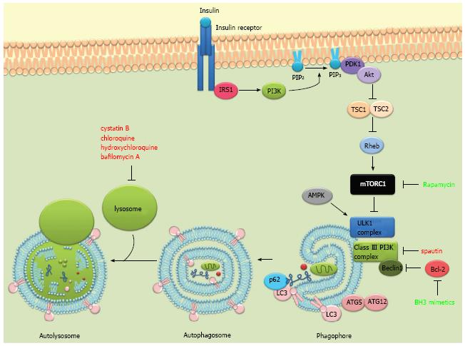
Figure 1 The autophagic process and some of its major regulators.
The phosphatidylinositol 3-kinase (PI3K) pathway is triggered by the binding of insulin or growth factors to the insulin receptor, activating PI3K. Activated PI3K converts PIP2 to PIP3 which then recruits PDK1 and Akt to the plasma membrane. Activated Akt then phosphorylates and inactivates TSC 1/2, leading to the activation of Rheb and mammalian target of rapamycin complex 1 (mTORC1). Under nutrient-rich conditions, mTORC1 suppresses autophagy by phosphorylating and inhibiting the ULK1 complex. Under starvation conditions or rapamycin treatment, mTOR dissociates from the ULK1 complex and autophagy gets activated. ULK1 is also directly phosphorylated and activated by the AMP-activated protein kinase (AMPK) in response to energy restriction. The autophagic process involves the degradation of cytosolic proteins and organelles in the lysosomes via autophagosomal delivery. Two complexes regulate the formation of the phagophore, the ULK1 complex and the beclin 1-VPS34 (class III PI3K) complex. The elongation of the autophagosomal membrane is mediated by two ubiquitin-like protein conjugation systems: ATG12-ATG5 and ATG8/LC3. LC3 can additionally recruit adaptor proteins such as p62 to autophagosomes mediating selective autophagy of cellular structures, protein aggregates and microorganisms. LC3II (LC3 bound to phosphatidylethanolamine) is recruited to both the inner and outer autophagosomal membrane. Autophagosomes fuse with lysosomes to form autolysosomes where they are degraded together with their luminal content. Pharmacological modulators of autophagy are labeled according to their effect on autophagy. Red: Inhibits autophagy; Green: Induces autophagy.
- Citation: Maycotte P, Thorburn A. Targeting autophagy in breast cancer. World J Clin Oncol 2014; 5(3): 224-240
- URL: https://www.wjgnet.com/2218-4333/full/v5/i3/224.htm
- DOI: https://dx.doi.org/10.5306/wjco.v5.i3.224


