Copyright
©2013 Baishideng Publishing Group Co.
World J Clin Oncol. Aug 10, 2013; 4(3): 70-74
Published online Aug 10, 2013. doi: 10.5306/wjco.v4.i3.70
Published online Aug 10, 2013. doi: 10.5306/wjco.v4.i3.70
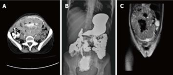
Figure 2 Results of image.
A: Computed tomography scan: oral and IV contrast revealed a large thick wall cavity mass (17 cm × 11 cm) with the enhancing wall; B: Barium follow-through delineated a large cavity connected to a segment of small bowel located at the mid-pelvic region; C: Magnetic resonance imaging of T1 and T2 coronal image shows a large cavity lesion containing gas and fluids and connected to a segment of small bowel but not related to the ovaries.
- Citation: Sawalhi S, Al-Harbi K, Raghib Z, Abdelrahman AI, Al-Hujaily A. Behavior of advanced gastrointestinal stromal tumor in a patient with von-Recklinghausen disease: Case report. World J Clin Oncol 2013; 4(3): 70-74
- URL: https://www.wjgnet.com/2218-4333/full/v4/i3/70.htm
- DOI: https://dx.doi.org/10.5306/wjco.v4.i3.70


