Copyright
©2012 Baishideng Publishing Group Co.
World J Clin Oncol. Jun 10, 2012; 3(6): 92-97
Published online Jun 10, 2012. doi: 10.5306/wjco.v3.i6.92
Published online Jun 10, 2012. doi: 10.5306/wjco.v3.i6.92
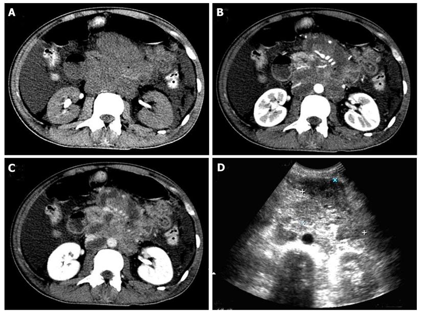
Figure 1 Abdominal computed tomography scan and ultrasonography examination revealed a cavitary mass between the distal jejunum and proximal ileum with multiple enlarged retroperitoneal lymph nodes.
A: Plain computed tomography (CT) scan image; B: CT image in the arterial phase of contrast enhancement; C: CT image in the parenchymal phase of contrast enhancement; D: Ultrasonography image.
- Citation: Li JZ, Tao J, Ruan DY, Yang YD, Zhan YS, Wang X, Chen Y, Kuang SC, Shao CK, Wu B. Primary duodenal NK/T-cell lymphoma with massive bleeding: A case report. World J Clin Oncol 2012; 3(6): 92-97
- URL: https://www.wjgnet.com/2218-4333/full/v3/i6/92.htm
- DOI: https://dx.doi.org/10.5306/wjco.v3.i6.92


