Copyright
©2011 Baishideng Publishing Group Co.
World J Clin Oncol. May 10, 2011; 2(5): 217-224
Published online May 10, 2011. doi: 10.5306/wjco.v2.i5.217
Published online May 10, 2011. doi: 10.5306/wjco.v2.i5.217
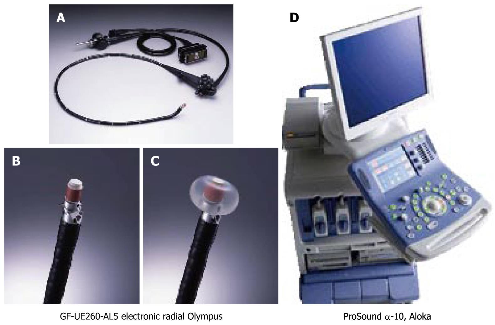
Figure 4 Endoscope and ultrasound machine (Setting-2).
A-C: The left three images indicate the whole view of the Olympus endosonoscope (GF-UE260-AL5) and the tip of the scope (both bare tip and tip with inflated balloon images are presented.); D: The image shows the ultrasound machine (ProSound α-10, Aloka, Japan).
- Citation: Hirooka Y, Itoh A, Kawashima H, Ohno E, Ishikawa T, Itoh Y, Nakamura Y, Hiramatsu T, Nakamura M, Miyahara R, Ohmiya N, Ishigami M, Katano Y, Goto H. Clinical oncology for pancreatic and biliary cancers: Advances and current limitations. World J Clin Oncol 2011; 2(5): 217-224
- URL: https://www.wjgnet.com/2218-4333/full/v2/i5/217.htm
- DOI: https://dx.doi.org/10.5306/wjco.v2.i5.217


