
















Copyright: ©Author(s) 2026.
World J Clin Oncol. Sep 24, 2026; 17(9): 125491
Published online Sep 24, 2026. doi: 10.5306/wjco.125491
Published online Sep 24, 2026. doi: 10.5306/wjco.125491
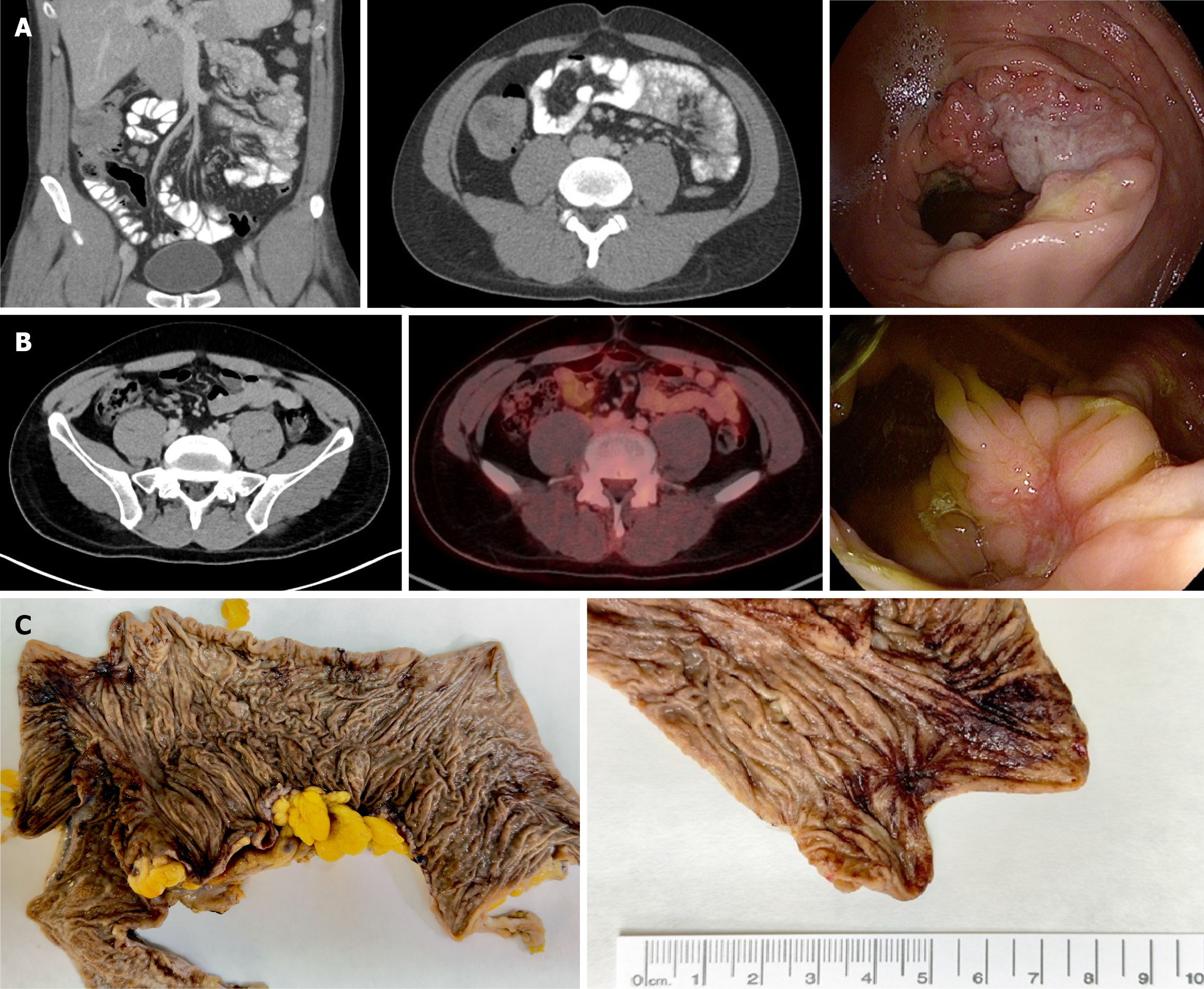
Figure 6 Case 5.
A: Coronal and axial abdominal computed tomography (CT) images and colonoscopy at diagnosis; B: Post-treatment abdominal CT, positron emission tomography/computed tomography, and colonoscopy after completion of neoadjuvant therapy; C: Gross pathology of the opened right hemicolectomy specimen showing an excavated lesion at the ileocecal junction extending into the ascending colon, corresponding to the treated tumor bed; detailed view of a 2 cm × 1.5 cm residual lesion at the post-treatment site.
- Citation: Manuel-Vazquez A, Soria Tristán M, Luján DR, Gortázar de las Casas S, Valle Rubio A, Ramos Rodríguez JL, García-Septiem J. Rethinking treatment for non-metastatic deficient mismatch repair colorectal and small bowel cancers. World J Clin Oncol 2026; 17(9): 125491
- URL: https://www.wjgnet.com/2218-4333/full/v17/i9/125491.htm
- DOI: https://dx.doi.org/10.5306/wjco.125491