
















Copyright: ©Author(s) 2026.
World J Clin Oncol. Sep 24, 2026; 17(9): 123454
Published online Sep 24, 2026. doi: 10.5306/wjco.123454
Published online Sep 24, 2026. doi: 10.5306/wjco.123454
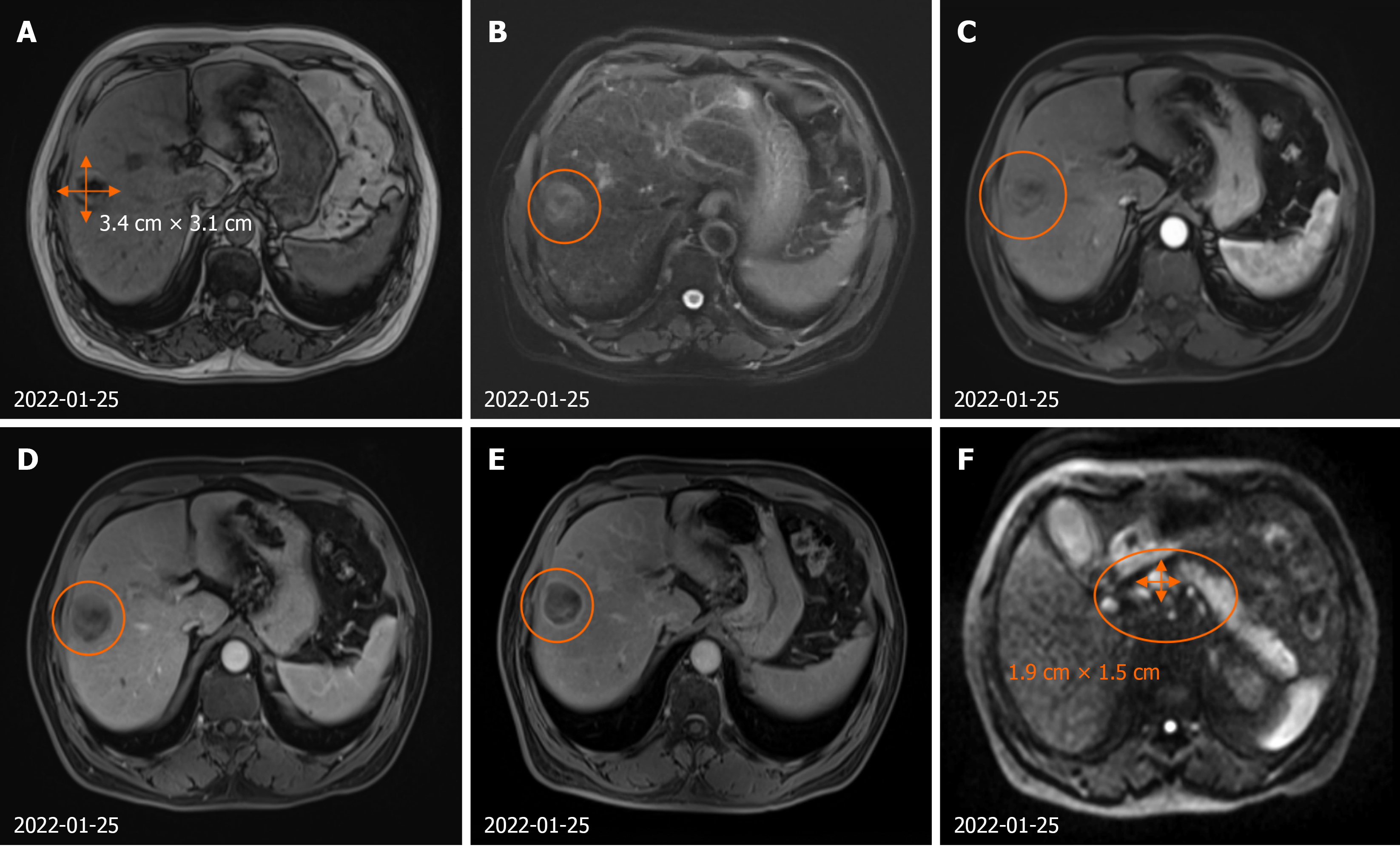
Figure 1 Hepatocellular carcinoma with multiple lymph node metastases in the liver, hepatic hilar region, peripancreatic head region, retroperitoneum, pelvic cavity, bilateral iliac vascular regions, and bilateral inguinal regions.
A: T1-weighted sequence of plain abdominal magnetic resonance imaging (MRI) at the junction of segments S8 and S5 of the liver; B: T2-weighted sequence of plain abdominal MRI at the junction of segments S8 and S5 of the liver; C: Arterial phase of enhanced abdominal MRI at the junction of segments S8 and S5 of the liver; D: Venous phase of enhanced abdominal MRI at the junction of segments S8 and S5 of the liver; E: Delayed phase of enhanced abdominal MRI at the junction of segments S8 and S5 of the liver; F: Multiple metastatic lymph nodes in the hepatic hilar region and peripancreatic head region.
- Citation: Yu SL, Liu JE, Li CY, Ling H, Tao HY, Shen YQ. Efficacy of transcatheter arterial embolization plus targeted chemotherapy for advanced hepatocellular carcinoma: A case report and literature review. World J Clin Oncol 2026; 17(9): 123454
- URL: https://www.wjgnet.com/2218-4333/full/v17/i9/123454.htm
- DOI: https://dx.doi.org/10.5306/wjco.123454