
















Copyright: ©Author(s) 2026.
World J Clin Oncol. Aug 24, 2026; 17(8): 124132
Published online Aug 24, 2026. doi: 10.5306/wjco.124132
Published online Aug 24, 2026. doi: 10.5306/wjco.124132
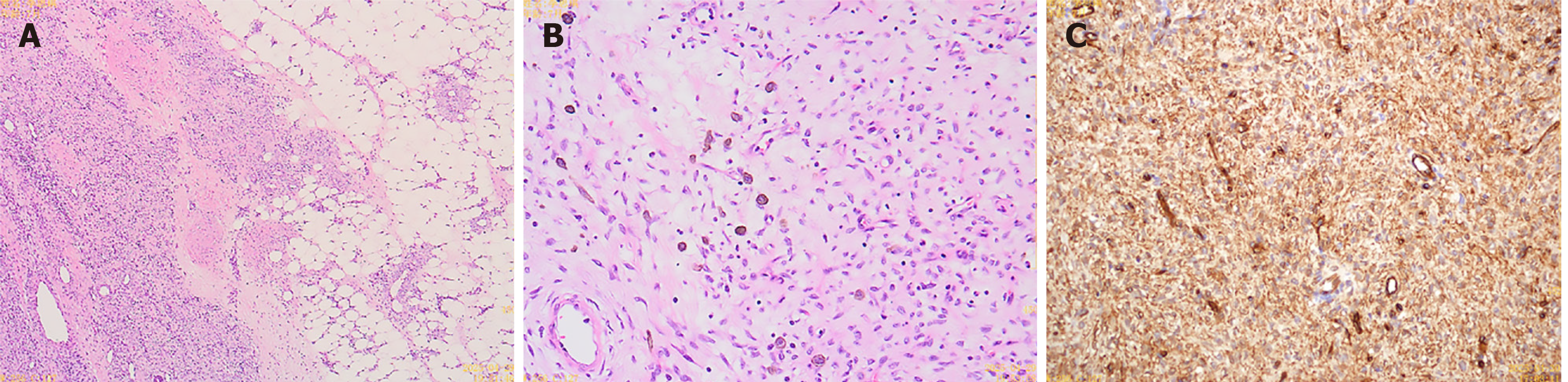
Figure 5 Final surgical specimen confirming classic dermatofibrosarcoma protuberans.
A: Low power hematoxylin and eosin image showing a dermal based spindle cell tumor with infiltrative extension into the subcutaneous adipose tissue in a characteristic honeycomb pattern (× 40 magnification, scale bar = 500 μm); B: Medium power hematoxylin and eosinimage demonstrating the classic storiform (cartwheel) arrangement of uniform spindle cells (× 100 magnification, scale bar = 200 μm); C: Immunohistochemical staining showing strong, diffuse positivity for CD34 antigen in the tumor cells (× 200 magnification, scale bar = 100 μm).
- Citation: Zheng CJ, Wan CL, Zhang CB, Hai B, Huang CF. Congenital dermatofibrosarcoma protuberans in a seven-month-old infant initially misdiagnosed as infantile myofibroma: A case report. World J Clin Oncol 2026; 17(8): 124132
- URL: https://www.wjgnet.com/2218-4333/full/v17/i8/124132.htm
- DOI: https://dx.doi.org/10.5306/wjco.124132