
















Copyright: ©Author(s) 2026.
World J Clin Oncol. Jun 24, 2026; 17(6): 120388
Published online Jun 24, 2026. doi: 10.5306/wjco.120388
Published online Jun 24, 2026. doi: 10.5306/wjco.120388
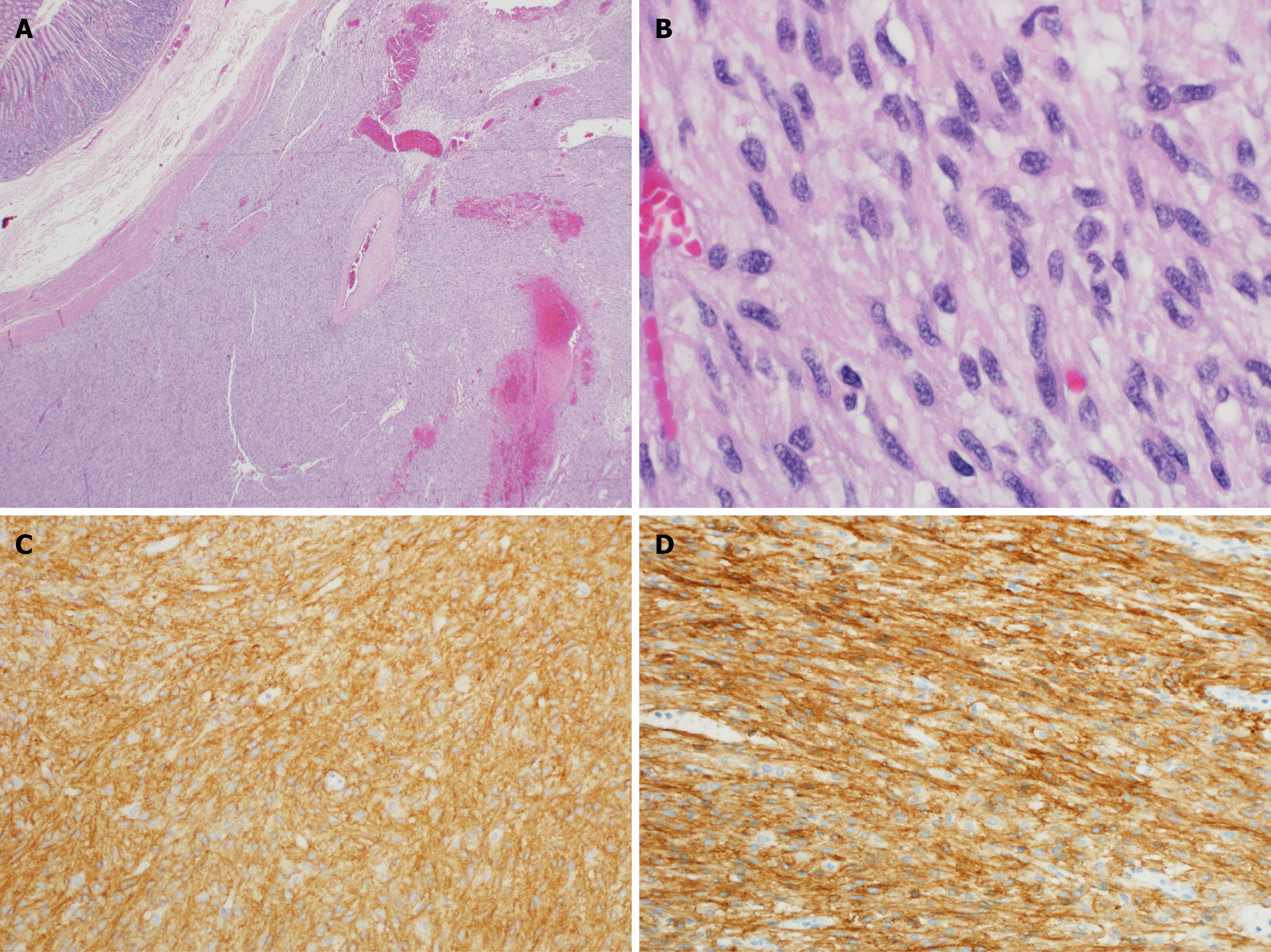
Figure 4 Example of microscopic view and immunohistochemistry of gastrointestinal stromal tumor.
A: Light microscopic view of gastrointestinal stromal tumor (GIST) tumors occupying the submucosa and muscularis propria, notice normal gastric mucosa in the left upper corner [hematoxylin and eosin (H&E) staining × 20]; B: High power light microscopic appearance of GIST tumor characterized by fascicles of spindle cells (H&E × 600); C: GIST tumor cells stain strongly and diffusely positive with DOG-1 antibody (Immunohistochemistry × 200); D: GIST tumor cells stain strongly and diffusely positive with CD117 antibody (Immunohistochemistry × 200).
- Citation: El-Matbouly MA, Al-Thani H, El-Menyar A, Al-Sulaiti M, Asim M, Al Yaseen M, Suliman A, Albahrani A, Al-Bozom I, Tabeb A. Evolving patterns of gastrointestinal stromal tumor presentation, management, and outcome over three decades: A Middle Eastern retrospective study. World J Clin Oncol 2026; 17(6): 120388
- URL: https://www.wjgnet.com/2218-4333/full/v17/i6/120388.htm
- DOI: https://dx.doi.org/10.5306/wjco.120388