
















Copyright: ©Author(s) 2026.
World J Clin Oncol. Jun 24, 2026; 17(6): 120388
Published online Jun 24, 2026. doi: 10.5306/wjco.120388
Published online Jun 24, 2026. doi: 10.5306/wjco.120388
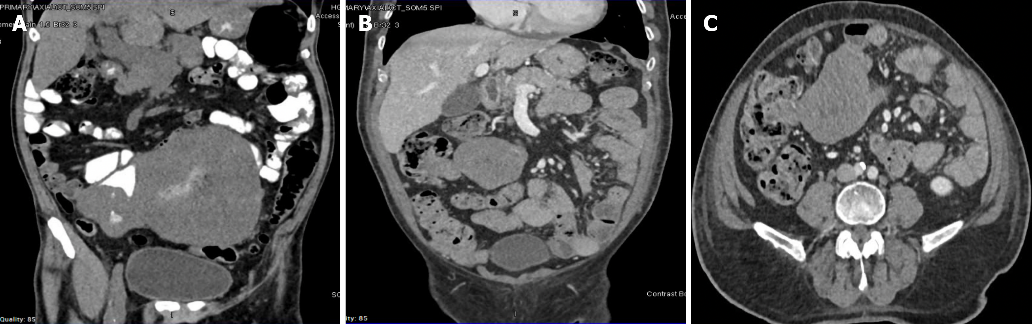
Figure 3 The radiological images of the small-bowel gastrointestinal stromal tumor.
A: A large heterogeneously enhancing mass measuring approximately 18.2 cm × 10.7 cm × 2.3 cm in the mid and lower abdomen, demonstrating hypodense areas and a large central area of necrosis. The mass appeared to arise/communicate with the loop of the ileum with oral contrast and air within the necrotic region; B: Radiodense soft-tissue mass arising from the jejunal loops in the right lower quadrant and hemipelvis; C: It measures 7.2 cm × 7.2 cm × 6.2 cm (maximum AP, transverse, and CC, respectively).
- Citation: El-Matbouly MA, Al-Thani H, El-Menyar A, Al-Sulaiti M, Asim M, Al Yaseen M, Suliman A, Albahrani A, Al-Bozom I, Tabeb A. Evolving patterns of gastrointestinal stromal tumor presentation, management, and outcome over three decades: A Middle Eastern retrospective study. World J Clin Oncol 2026; 17(6): 120388
- URL: https://www.wjgnet.com/2218-4333/full/v17/i6/120388.htm
- DOI: https://dx.doi.org/10.5306/wjco.120388