
















Copyright: ©Author(s) 2026.
World J Clin Oncol. Jun 24, 2026; 17(6): 120388
Published online Jun 24, 2026. doi: 10.5306/wjco.120388
Published online Jun 24, 2026. doi: 10.5306/wjco.120388
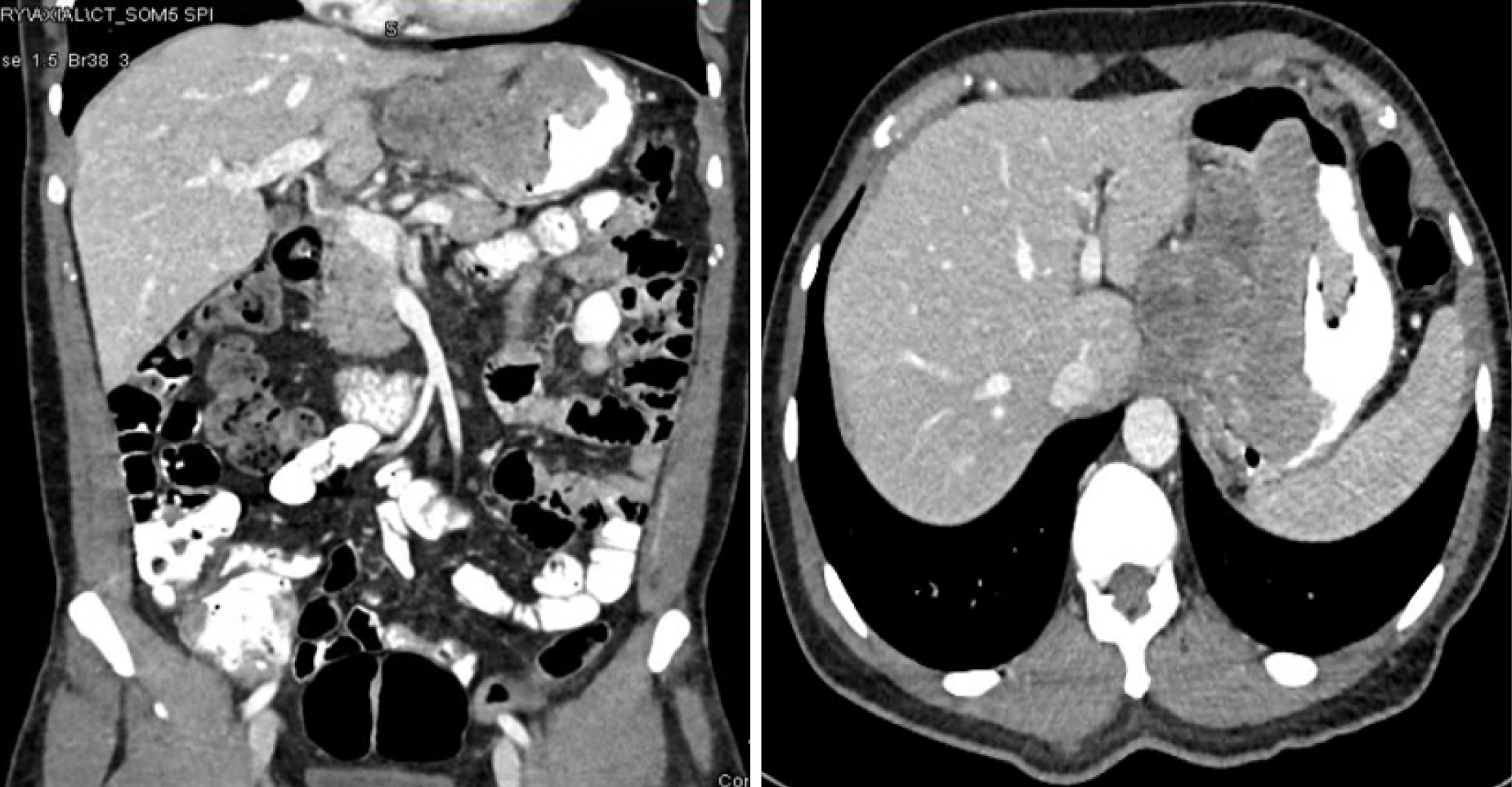
Figure 2 Coronal and cross-sectional computed tomography scan cuts of gastric gastrointestinal stromal tumor, showing the exophytic component of the gastric tumor with proximity to the left lobe of the liver.
Computed tomography scans showing gastric gastrointestinal stromal tumor: A large heterogeneous hypodense lobulated partly exophytic mass measuring about 10.5 cm in maximal dimension was noted at the gastric fundus, showing ulceration of its intraluminal component; superiorly, it abutted the left hemidiaphragm (probable infiltrate) and the lateral aspect of the left hepatic lobe.
- Citation: El-Matbouly MA, Al-Thani H, El-Menyar A, Al-Sulaiti M, Asim M, Al Yaseen M, Suliman A, Albahrani A, Al-Bozom I, Tabeb A. Evolving patterns of gastrointestinal stromal tumor presentation, management, and outcome over three decades: A Middle Eastern retrospective study. World J Clin Oncol 2026; 17(6): 120388
- URL: https://www.wjgnet.com/2218-4333/full/v17/i6/120388.htm
- DOI: https://dx.doi.org/10.5306/wjco.120388