
















Copyright: ©Author(s) 2026.
World J Clin Oncol. May 24, 2026; 17(5): 119615
Published online May 24, 2026. doi: 10.5306/wjco.v17.i5.119615
Published online May 24, 2026. doi: 10.5306/wjco.v17.i5.119615
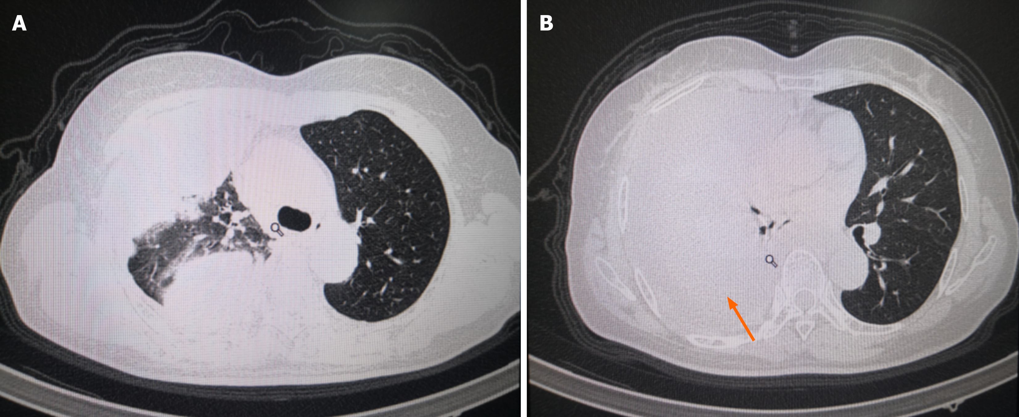
Figure 5 Radiologic progression during sorafenib therapy.
A and B: A computed tomography scan obtained in February 2022, following several months of firstline sorafenib treatment, shows extensive bilateral pulmonary metastases and a substantial increase in the rightsided pleural effusion compared with prior imaging (arrows). These findings, together with rising serum thyroglobulin levels and worsening respiratory symptoms, indicate progressive disease under sorafenib.
- Citation: Zhou CY, Ren XY, Li HH, Pan XM, Lan X, Duan D. Anlotinib plus albumin-bound paclitaxel induction-maintenance for sorafenib-refractory radioiodine-refractory differentiated thyroid cancer: A case report and review of literature. World J Clin Oncol 2026; 17(5): 119615
- URL: https://www.wjgnet.com/2218-4333/full/v17/i5/119615.htm
- DOI: https://dx.doi.org/10.5306/wjco.v17.i5.119615