
















Copyright: ©Author(s) 2026.
World J Clin Oncol. May 24, 2026; 17(5): 119443
Published online May 24, 2026. doi: 10.5306/wjco.v17.i5.119443
Published online May 24, 2026. doi: 10.5306/wjco.v17.i5.119443
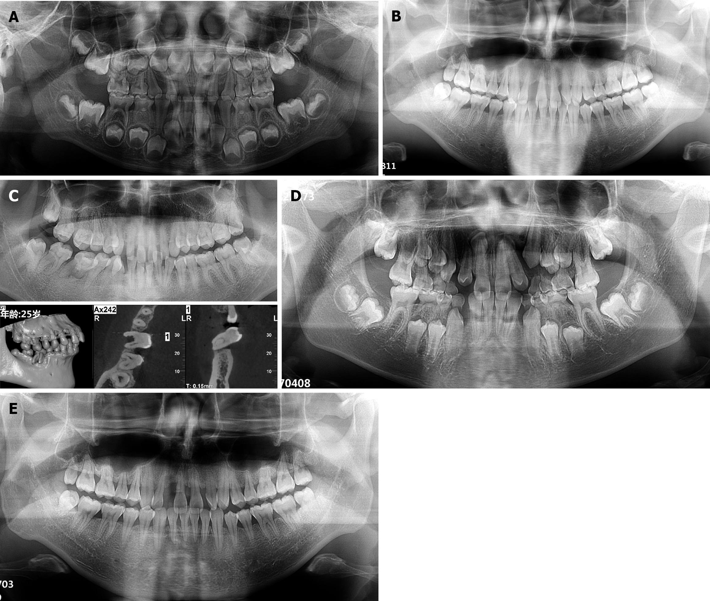
Figure 7 Imaging findings.
A: Orthopantomogram (OPG) of patient 1 showing all permanent tooth buds present without alveolar bone loss; B: OPG of patient 2 showing extensive alveolar bone resorption, particularly in the maxillary anterior region; C: OPG and cone-beam computed tomography of patient 3 showing # 46 in linguoclination with severe alveolar bone resorption; D: OPG of patient 4 showing mixed dentition without alveolar bone loss; permanent teeth # 12-22, 32-42, 16, 26, 36, and 46 had erupted, while deciduous teeth # 53-55, 63-65, 74, 75, 84 and 85 were still present; E: OPG of patient 2 at 3-year follow-up after periodontal surgery showing no further alveolar bone loss.
- Citation: Cheng Y, Zhao J, Yang J, Lin LY, Wu J. Clinical presentation and management of hereditary gingival fibromatosis: Four case reports. World J Clin Oncol 2026; 17(5): 119443
- URL: https://www.wjgnet.com/2218-4333/full/v17/i5/119443.htm
- DOI: https://dx.doi.org/10.5306/wjco.v17.i5.119443