
















Copyright: ©Author(s) 2026.
World J Clin Oncol. May 24, 2026; 17(5): 118350
Published online May 24, 2026. doi: 10.5306/wjco.v17.i5.118350
Published online May 24, 2026. doi: 10.5306/wjco.v17.i5.118350
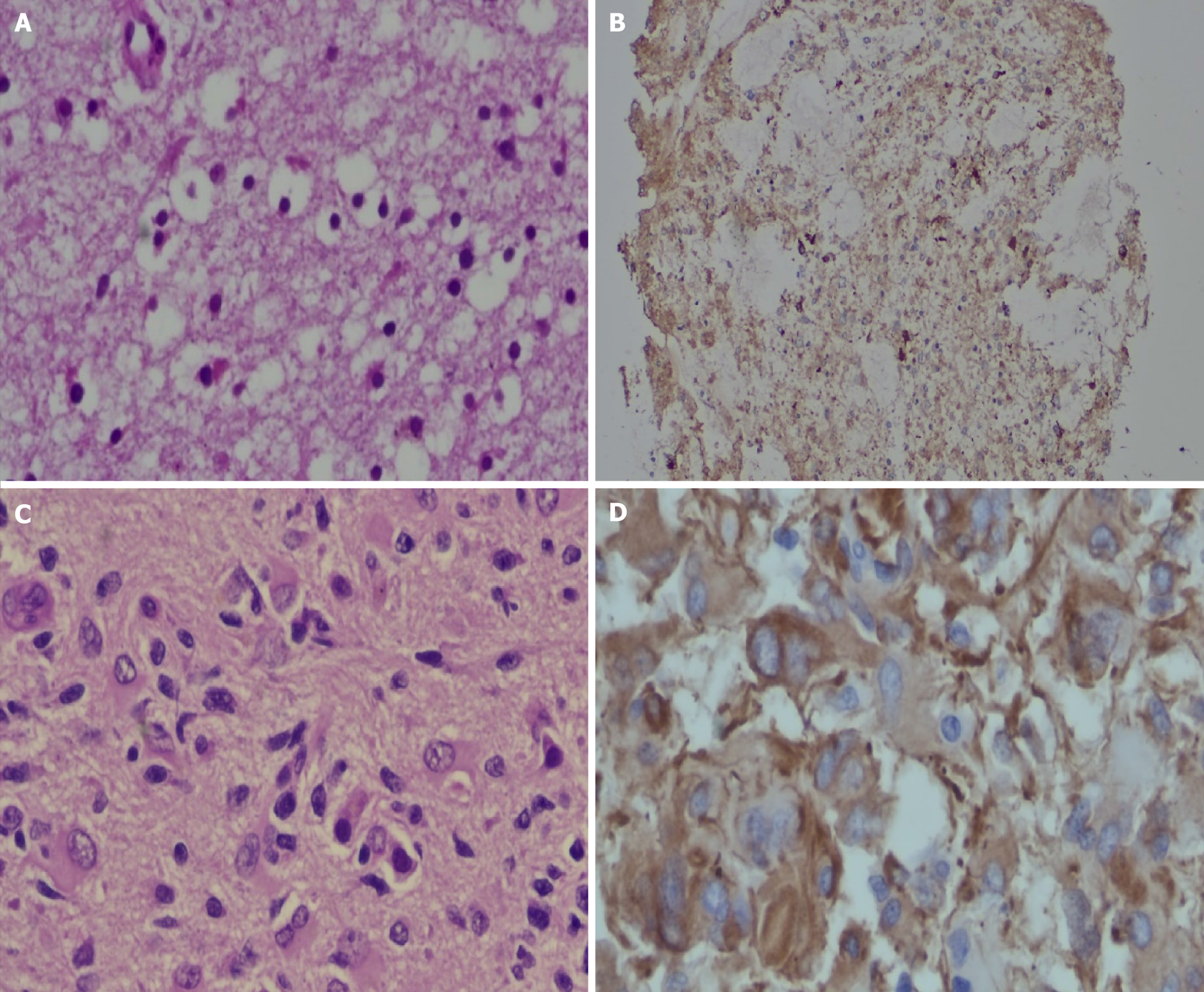
Figure 1 Representative images of morphology and immunohistochemical staining of glioma tumors.
A: Low-grade astrocytoma (World Health Organization grade II) on hematoxylin and eosin [hematoxylin-eosin (HE)] staining showing low-grade and uniform nuclei (HE, × 100); B: Cytoplasmic nestin expression on immunohistochemistry (IHC) staining in a low-grade glioma case (IHC for nestin, × 50); C: High-grade glioblastoma (World Health Organization grade IV) on HE staining with high-grade pleomorphic nuclei (HE, × 200); D: Strong cytoplasmic nestin expression on IHC staining in a high-grade glioma case (IHC for nestin, × 200).
- Citation: Nasir S, Ali A, Ullah I, Wazir S, Javaid I, Jalal SZ, Khan I, Mubarak M. Clinicopathological and prognostic relevance of nestin in low- and high-grade gliomas: An immunohistochemical study. World J Clin Oncol 2026; 17(5): 118350
- URL: https://www.wjgnet.com/2218-4333/full/v17/i5/118350.htm
- DOI: https://dx.doi.org/10.5306/wjco.v17.i5.118350