
















Copyright: ©Author(s) 2026.
World J Clin Oncol. Mar 24, 2026; 17(3): 117055
Published online Mar 24, 2026. doi: 10.5306/wjco.v17.i3.117055
Published online Mar 24, 2026. doi: 10.5306/wjco.v17.i3.117055
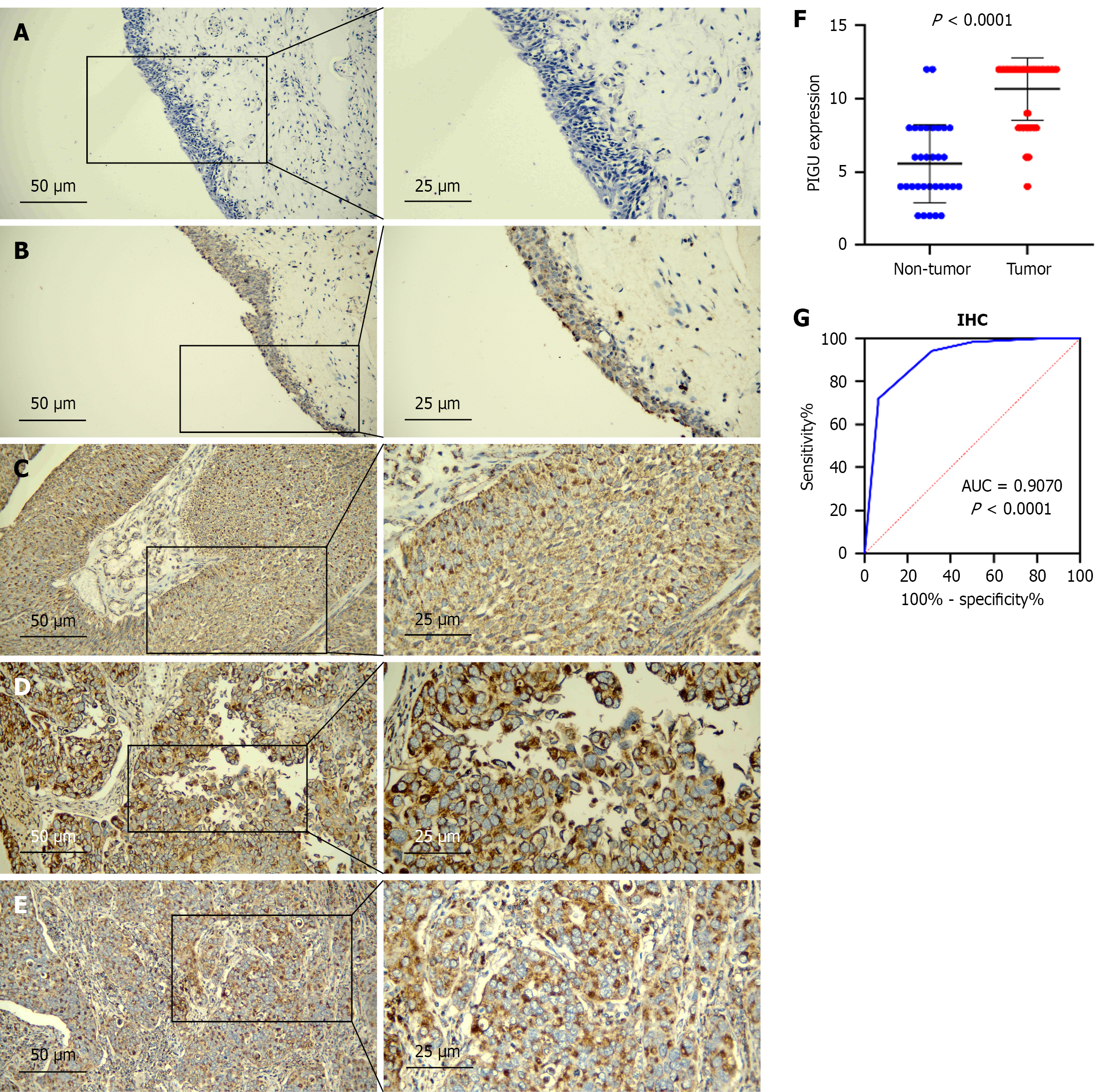
Figure 1 Immunohistochemistry detection of PIGU protein expression in bladder cancer tissues.
PIGU protein expression in para-cancerous tissues. A: Negative, × 200 (left), × 400 (right); B: Weakly positive, × 200 (left), × 400 (right); C-E: PIGU protein expression in bladder cancer (BLCA) tissue, all 12 points, × 200 (left), × 400 (right), pathological grade I-III (C: PIGU protein expression in BLCA tissue, positive, × 200-left, × 400-right); F: Scatter plot of immunohistochemical results; G: Receiver operating characteristic curve. IHC: Immunohistochemistry.
- Citation: Li SH, Liu ZS, Chen YJ, Li JD, Wei L, Chen YY, Zhang W, Huang ZG, Chen ZD, Chen G, Wei DM, Mo ZN, Deng LL. PIGU overexpression and regulation of cellular biological functions in bladder cancer. World J Clin Oncol 2026; 17(3): 117055
- URL: https://www.wjgnet.com/2218-4333/full/v17/i3/117055.htm
- DOI: https://dx.doi.org/10.5306/wjco.v17.i3.117055