
















Copyright: ©Author(s) 2026.
World J Clin Oncol. Mar 24, 2026; 17(3): 114744
Published online Mar 24, 2026. doi: 10.5306/wjco.v17.i3.114744
Published online Mar 24, 2026. doi: 10.5306/wjco.v17.i3.114744
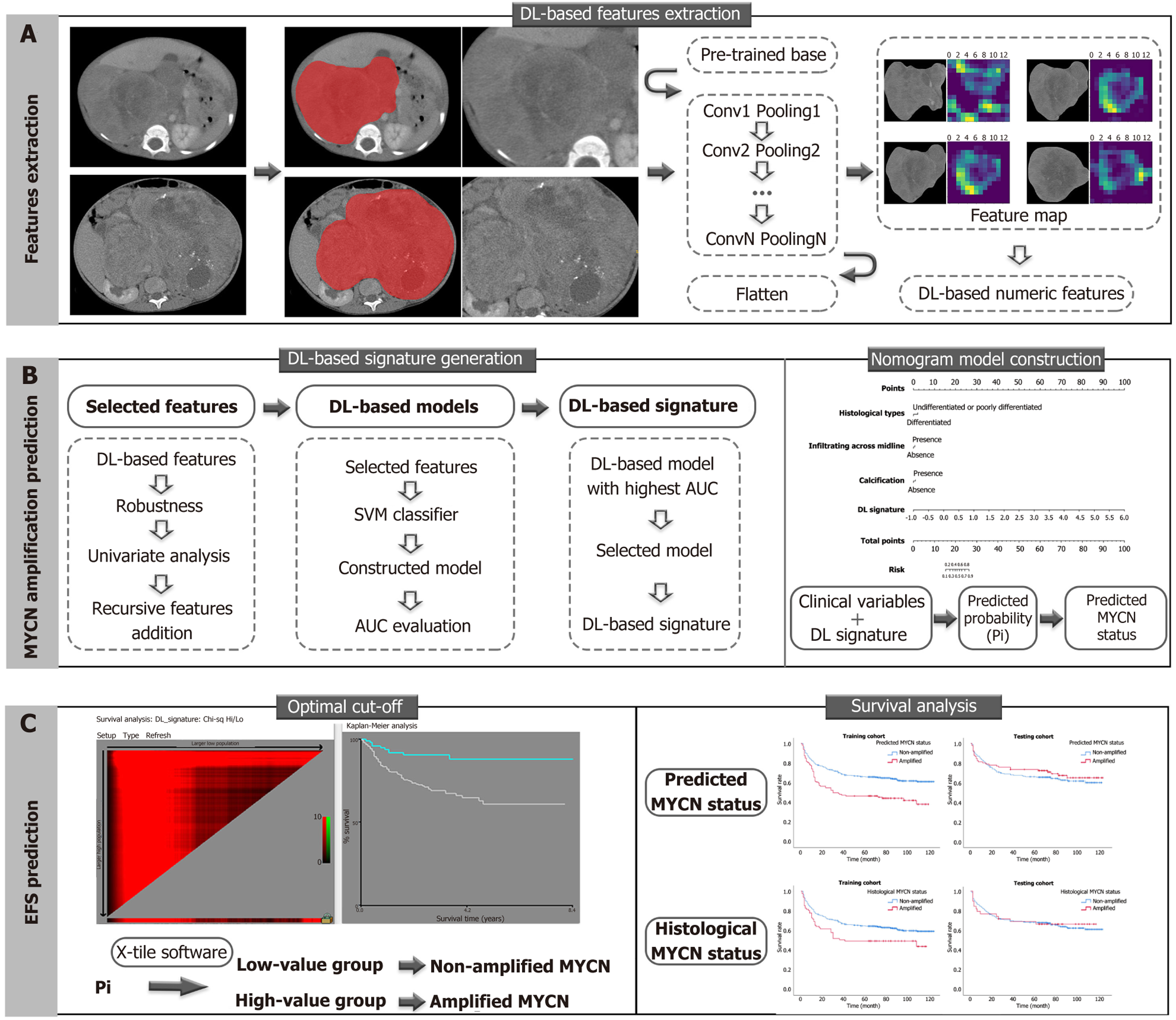
Figure 1 A general flowchart of data analysis.
A: Deep learning (DL)-based feature extraction was performed on contrast-enhanced computed tomography images of neuroblastoma by pre-trained convolutional neural network algorithms, which transformed the original images into numeric DL-based features; B: The MYCN amplification prediction was approached by DL-based features via DL-based signature generation and integrated nomogram model construction. The DL-based signature was generated on the DL-based model with the largest area under the receiver operating characteristic curve value in the testing cohort. The integrated nomogram model combined significant clinical variables and the DL-based signature; C: The survival analysis in the prediction of event-free survival was evaluated on MYCN amplification identified by nomogram-predicted and histopathological results. Predicted probabilities (Pi) are the total points that derive from independent variables in the nomogram model for differentiating non-amplified and amplified MYCN status. The optimal cut-off point of Pi was -1.996, defined by X-tile software, which indicated Pi ≤ -1.996 is considered as non-amplified MYCN status, and Pi > -1.996 is considered as amplified MYCN status. DL: Deep learning; SVM: Support vector machine; AUC: Area under the receiver operating characteristic curve; EFS: Event-free survival; Pi: Predicted probabilities.
- Citation: Yang YH, Li Y. Deep learning radiomic analysis in the prediction of MYCN status and survival outcome in children with neuroblastoma. World J Clin Oncol 2026; 17(3): 114744
- URL: https://www.wjgnet.com/2218-4333/full/v17/i3/114744.htm
- DOI: https://dx.doi.org/10.5306/wjco.v17.i3.114744