
















©The Author(s) 2026.
World J Clin Oncol. Feb 24, 2026; 17(2): 113113
Published online Feb 24, 2026. doi: 10.5306/wjco.v17.i2.113113
Published online Feb 24, 2026. doi: 10.5306/wjco.v17.i2.113113
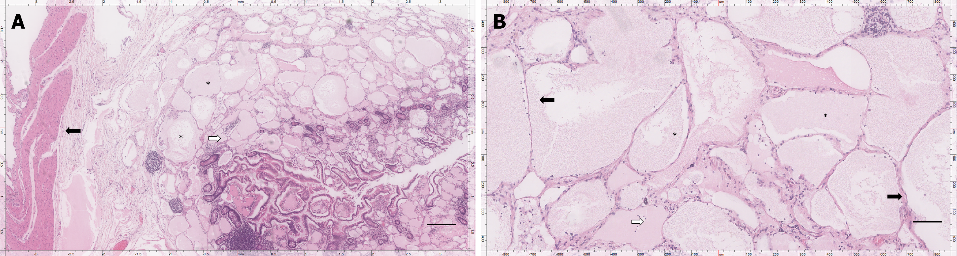
Figure 3 Histopathological examination of the resected small bowel mass confirming lymphatic malformation.
A: Low power magnification (× 10). Small bowel with multiple dilated, thin-walled lymphatic channels within the submucosa and muscularis layers. The cystic spaces are lined with flattened endothelial cells and contain proteinaceous fluid, consistent with lymphatic malformation (hematoxylin and eosin stain). Scale bar = 100 μm; B: High power magnification (× 40). Lymphatic malformation of the small bowel. Seen are multiple, thin-walled endothelial-lined cystic spaces filled with pale eosinophilic fluid, separated by delicate fibrous septa, with scattered lymphocytes (hematoxylin and eosin stain). Scale bar = 50 μm. Stars (A, B): Cystically dilated lymphatic channels with pale eosinophilic proteinaceous material in lumen.
- Citation: Khalayleh H, Bader R, Abu Arafeh M, Odeh Q, Rogalsky B, Imam R, Khalaileh A, Imam A. Small bowel lymphatic malformation: Clinical presentation and a comprehensive literature review. World J Clin Oncol 2026; 17(2): 113113
- URL: https://www.wjgnet.com/2218-4333/full/v17/i2/113113.htm
- DOI: https://dx.doi.org/10.5306/wjco.v17.i2.113113