
















©The Author(s) 2026.
World J Clin Oncol. Feb 24, 2026; 17(2): 113113
Published online Feb 24, 2026. doi: 10.5306/wjco.v17.i2.113113
Published online Feb 24, 2026. doi: 10.5306/wjco.v17.i2.113113
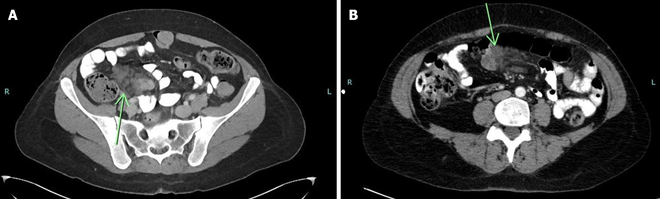
Figure 1 Axial contrast-enhanced computed tomography scans of the abdomen showing small bowel lymphatic malformation.
A: A lobulated, low-attenuation lesion within the mesentery adjacent to small bowel loops (green arrow), suggestive of a lymphatic malformation; B: The lesion appears to contain internal septations and shows no evidence of enhancement (green arrow), consistent with the imaging features of a mesenteric lymphatic malformation.
- Citation: Khalayleh H, Bader R, Abu Arafeh M, Odeh Q, Rogalsky B, Imam R, Khalaileh A, Imam A. Small bowel lymphatic malformation: Clinical presentation and a comprehensive literature review. World J Clin Oncol 2026; 17(2): 113113
- URL: https://www.wjgnet.com/2218-4333/full/v17/i2/113113.htm
- DOI: https://dx.doi.org/10.5306/wjco.v17.i2.113113