
















Copyright: ©Author(s) 2026.
World J Clin Oncol. Nov 24, 2026; 17(11): 124935
Published online Nov 24, 2026. doi: 10.5306/wjco.124935
Published online Nov 24, 2026. doi: 10.5306/wjco.124935
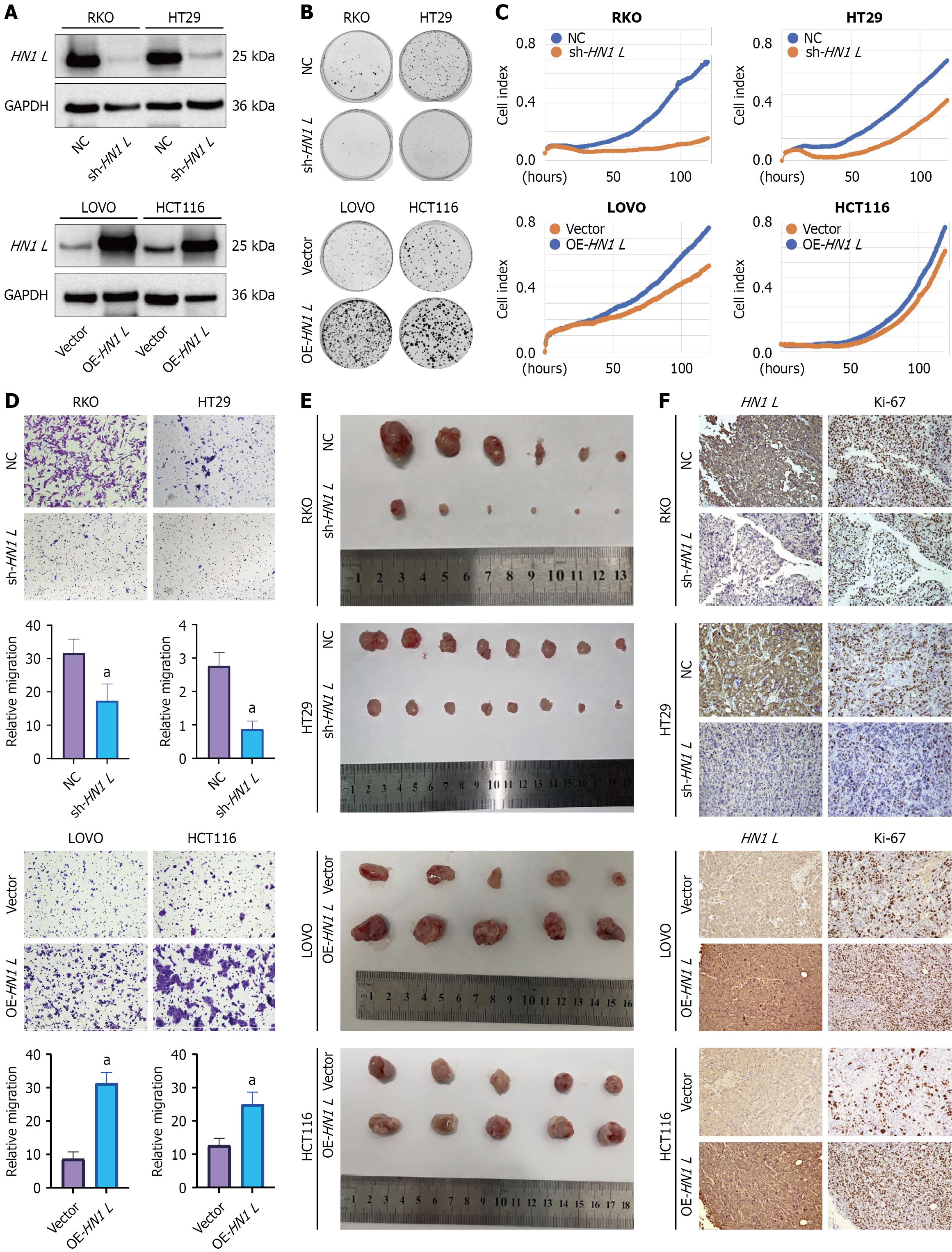
Figure 2 HN1 L promotes colorectal cancer tumorigenic potential in vitro and in vivo.
A: Western blot confirming HN1 L overexpression and sh-HN1 L#2-mediated knockdown in colorectal cancer (CRC) cells; B: Representative images and quantification of colony formation assays in HN1 L-modified CRC cells; C: Real-time proliferation curves measured using the IncuCyte system. The cell index represents the proportion of the image area occupied by cells; D: Representative images and quantification of Transwell migration assays; E: Representative endpoint images of subcutaneous xenograft tumors generated from CRC cells with HN1 L overexpression or knockdown; F: Representative immunohistochemical staining of HN1 L and Ki-67 in xenograft tissues. Quantitative data are presented as mean ± SD from at least three independent experiments. aP < 0.01 vs the corresponding control. OE: Overexpression; NC: Negative control.
- Citation: Kang D, Zeng LX, Zhang RX, Ma WJ, Yang R, Wang X. HN1 L promotes colorectal cancer proliferation and migration by inhibiting the Hippo pathway. World J Clin Oncol 2026; 17(11): 124935
- URL: https://www.wjgnet.com/2218-4333/full/v17/i11/124935.htm
- DOI: https://dx.doi.org/10.5306/wjco.124935