
















Copyright: ©Author(s) 2026.
World J Clin Oncol. Nov 24, 2026; 17(11): 124935
Published online Nov 24, 2026. doi: 10.5306/wjco.124935
Published online Nov 24, 2026. doi: 10.5306/wjco.124935
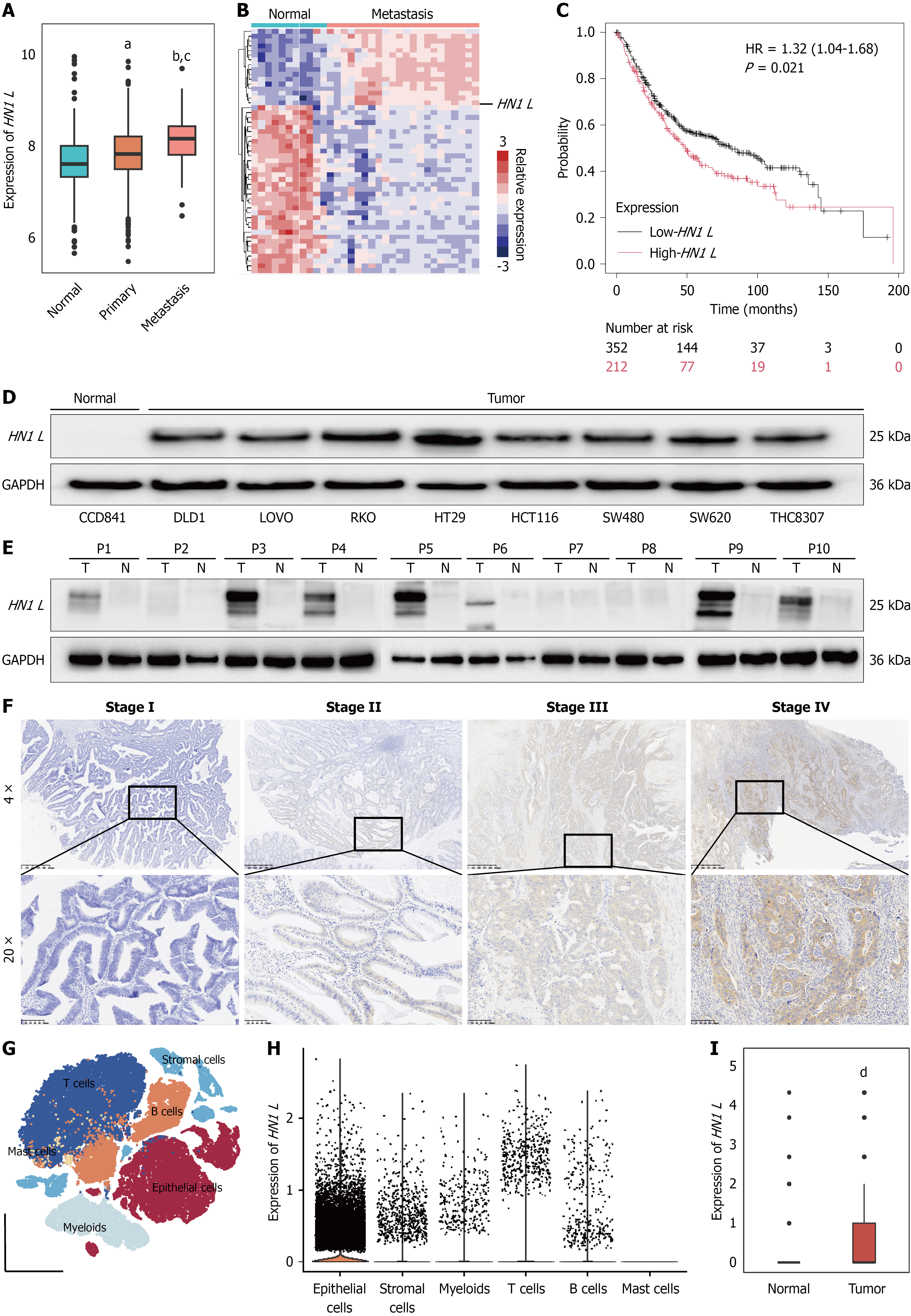
Figure 1 Increased expression of HN1 L in colorectal cancer.
A: HN1 L expression in normal colorectal tissues, primary colorectal cancer (CRC) tissues, and metastatic tumors from The Cancer Genome Atlas-CRC dataset; B: Heatmap showing HN1 L and representative differentially expressed genes in normal tissues and metastatic CRC tissues; C: Kaplan-Meier analysis of overall survival according to HN1 L expression in CRC; D: Western blot analysis of basal HN1 L expression in the normal colonic epithelial cell line CCD841 and eight CRC cell lines; E: Western blot analysis of HN1 L expression in an independent set of 10 paired frozen CRC and adjacent normal tissues; F: Representative immunohistochemical staining of HN1 L in CRC specimens from patients with different tumor stages; G: T-SNE visualization of the major cell populations identified in the GSE132465 scRNA-seq dataset; H and I: Distribution of HN1 L expression across major cell populations and tissues. ᵃP < 0.05, ᵇP < 0.01 vs normal tissues; ᶜP < 0.05 vs primary colorectal cancer tissues; dP < 0.01 vs normal tissues. HR: Hazard ratio.
- Citation: Kang D, Zeng LX, Zhang RX, Ma WJ, Yang R, Wang X. HN1 L promotes colorectal cancer proliferation and migration by inhibiting the Hippo pathway. World J Clin Oncol 2026; 17(11): 124935
- URL: https://www.wjgnet.com/2218-4333/full/v17/i11/124935.htm
- DOI: https://dx.doi.org/10.5306/wjco.124935