
















Copyright: ©Author(s) 2026.
World J Clin Oncol. Nov 24, 2026; 17(11): 124859
Published online Nov 24, 2026. doi: 10.5306/wjco.124859
Published online Nov 24, 2026. doi: 10.5306/wjco.124859
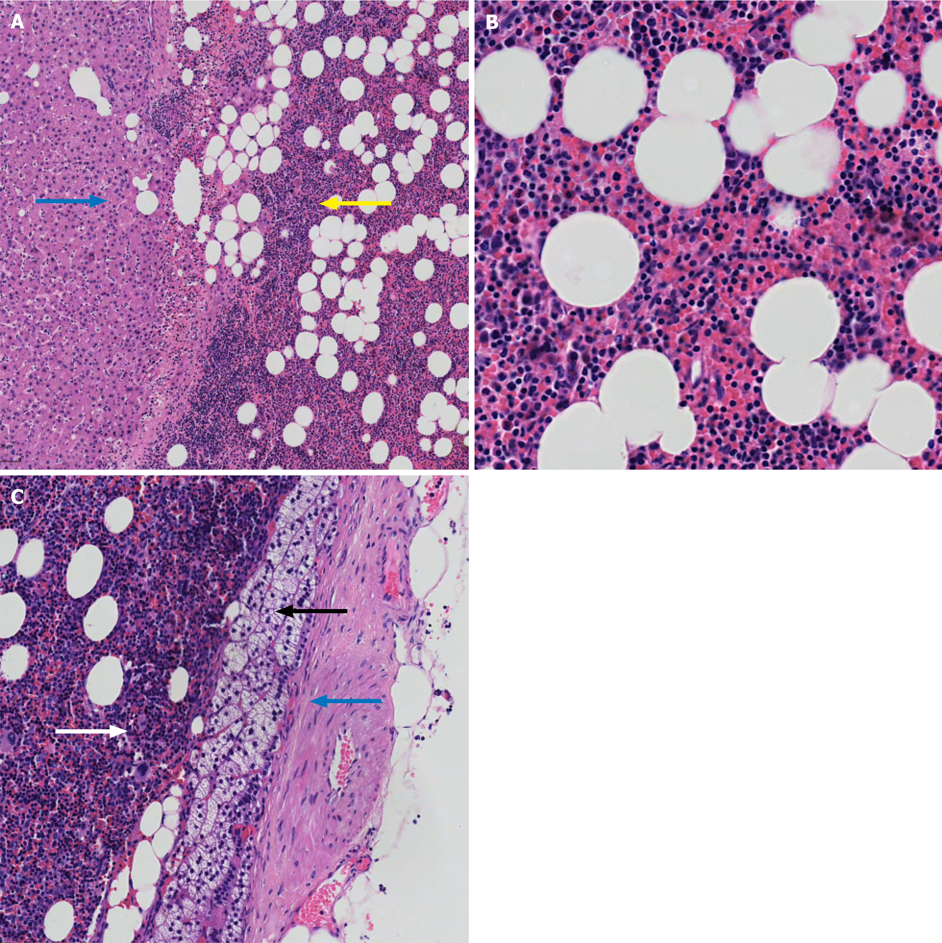
Figure 3 Microscopic views of resected specimens with hematoxylin and eosin staining.
A: Normal liver parenchyma (blue arrows) and myelolipoma (yellow arrows), 10 × magnification; B: The myelolipoma comprises adipose and hematopoietic tissues, including megakaryocytes and myelopoiesis, 40 × magnification; C: The tumor is observed within the capsule (blue arrows) consisting of adipose and myelopoietic tissues (white arrow), with a small amount of adrenal tissue (black arrow) in the immediate inner area of the capsule; 20 × magnification.
- Citation: Su CS, Zhu XY, Li H, Zhang SG. Ectopic adrenal myelolipoma at the hepatic-adrenal interface mimicking hepatic angiomyolipoma: A case report. World J Clin Oncol 2026; 17(11): 124859
- URL: https://www.wjgnet.com/2218-4333/full/v17/i11/124859.htm
- DOI: https://dx.doi.org/10.5306/wjco.124859