
















Copyright: ©Author(s) 2026.
World J Clin Oncol. Oct 24, 2026; 17(10): 126647
Published online Oct 24, 2026. doi: 10.5306/wjco.126647
Published online Oct 24, 2026. doi: 10.5306/wjco.126647
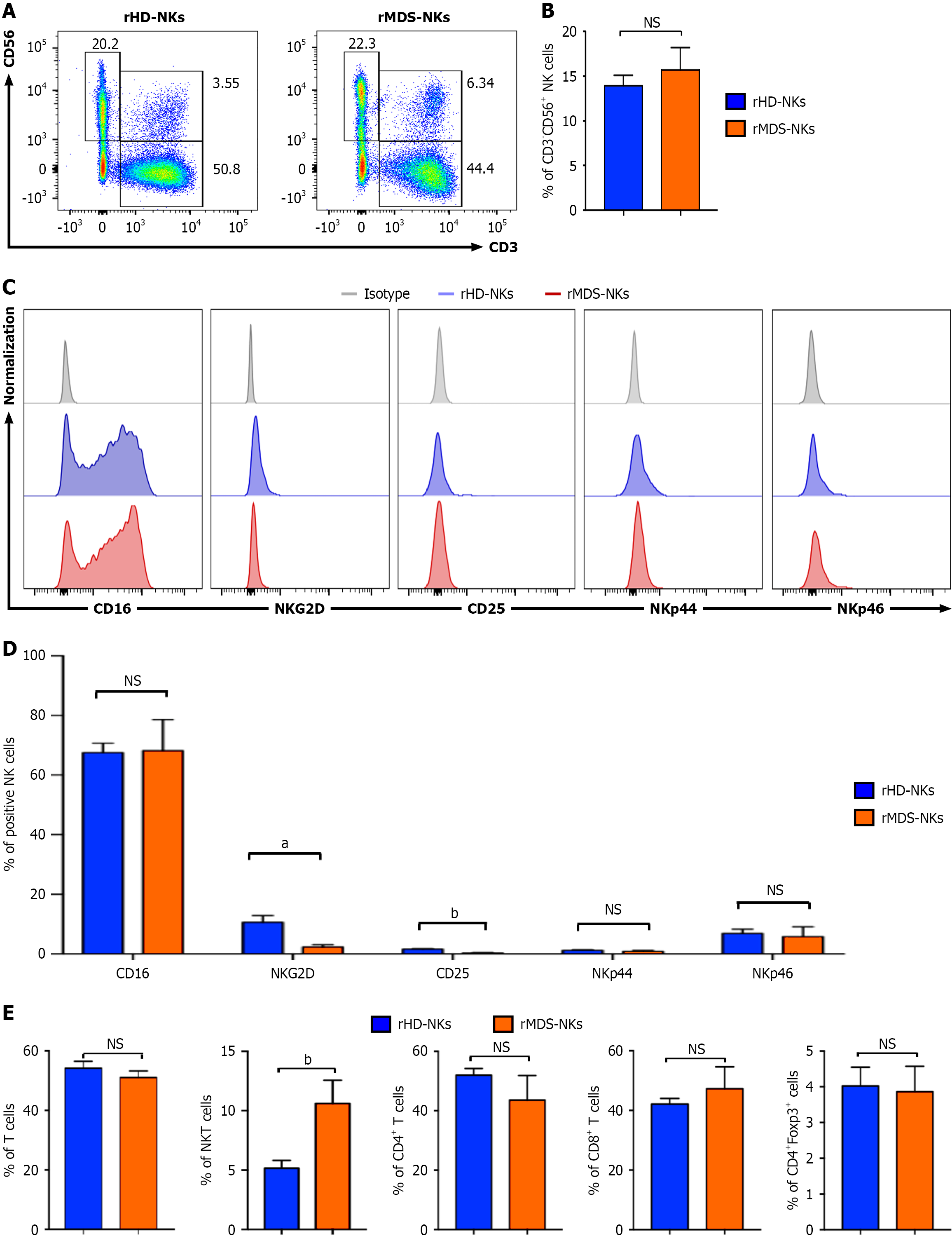
Figure 1 Comparison of the immunophenotype and components between resident myelodysplastic syndrome natural killers and resident healthy donor natural killers.
A and B: Representative flow cytometry diagrams (A) and statistical analysis (B) of CD3-CD56+ total natural killers (NK) cells in the indicated groups [resident myelodysplastic syndrome NKs (rMDS-NKs), resident healthy donor NKs (rHD-NKs)]; C and D: Representative flow cytometry diagrams (C) and statistical analyses (D) of total activated CD3-CD56+CD16+ NK cells and the indicated subsets (NKG2D+, CD25+, NKp44+, and NKp46+) in the indicated groups (rMDS-NKs, rHD-NKs); E: Statistical analyses of CD3+ total T lymphocytes, CD3+CD56+ NKT cells, and the T cell subsets (CD3+CD4+, CD3+CD8+, and CD4+FoxP3+ T cells) in the indicated groups (rMDS-NKs, rHD-NKs). Data were shown as mean ± SEM (n = 3 repetitions for each sample). aP < 0.05, bP < 0.01, NS: Not significant. rMDS-NKs: Resident myelodysplastic syndrome natural killers; rHD-NKs: Resident healthy donor natural killers; NK: Natural killer.
- Citation: Liu SY, Feng XM, Chen ZY, Chen ZQ, Shang YR, Linnebacher M, Wang YX, Zhang SS, Cai M, Zhang LS. Characterization of the cellular and transcriptomic landscapes of natural killer cells in hematopoietic microenvironment of myelodysplastic syndrome. World J Clin Oncol 2026; 17(10): 126647
- URL: https://www.wjgnet.com/2218-4333/full/v17/i10/126647.htm
- DOI: https://dx.doi.org/10.5306/wjco.126647