
















©The Author(s) 2026.
World J Clin Oncol. Jan 24, 2026; 17(1): 113966
Published online Jan 24, 2026. doi: 10.5306/wjco.v17.i1.113966
Published online Jan 24, 2026. doi: 10.5306/wjco.v17.i1.113966
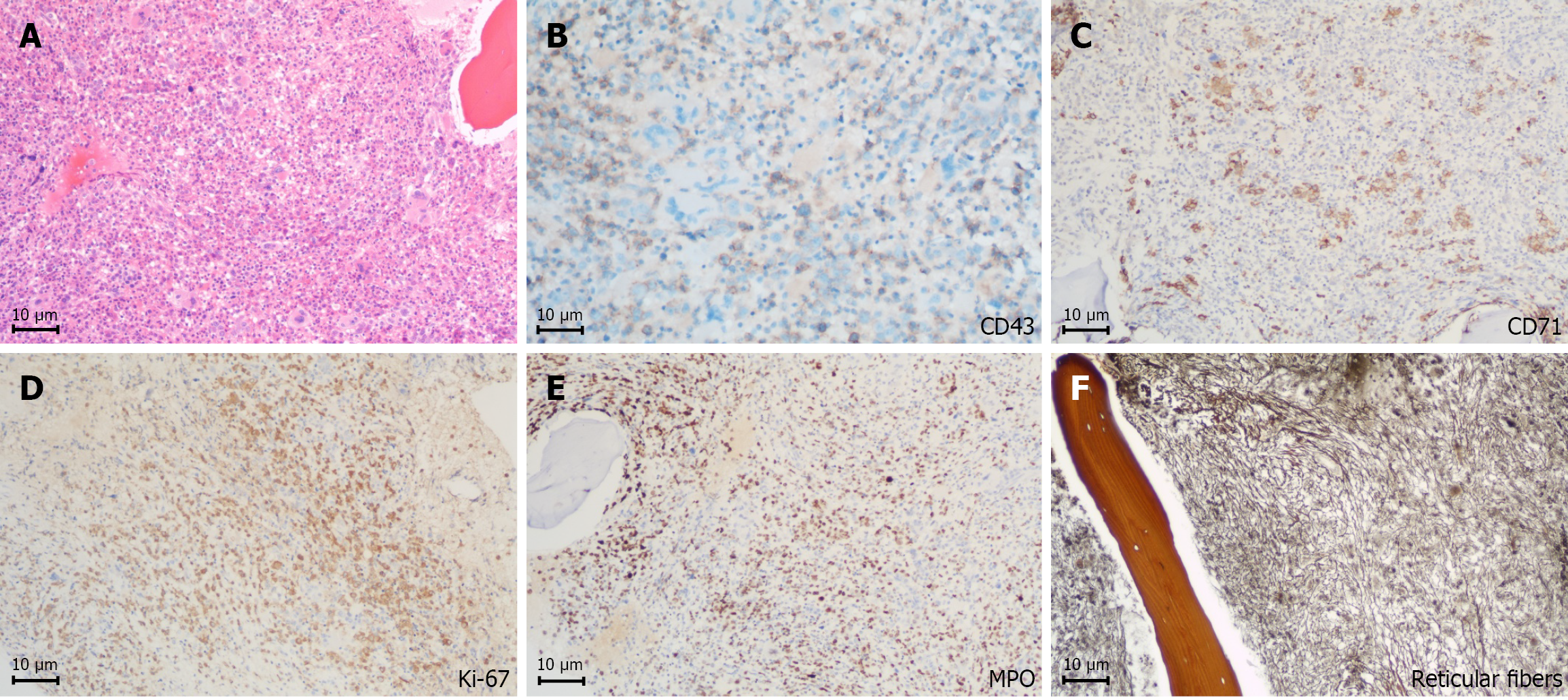
Figure 1 Pathological results.
A: Hematoxylin-eosin staining of the left femoral head section showing the pathological lesion; B-E: Histopathology of myeloid sarcoma with positive staining for CD42b (B), CD71 (C), Ki-67 (D), and myeloperoxidase (E); F: Reticular fiber staining in the bone marrow (magnification × 10).
- Citation: Zhong SH, Lou J, Zhao AQ, Jin XL, Wei SM, Liang Y. Myeloid sarcoma transformed from myeloproliferative neoplasm: A case report and review of literature. World J Clin Oncol 2026; 17(1): 113966
- URL: https://www.wjgnet.com/2218-4333/full/v17/i1/113966.htm
- DOI: https://dx.doi.org/10.5306/wjco.v17.i1.113966