
















©The Author(s) 2026.
World J Clin Oncol. Jan 24, 2026; 17(1): 111426
Published online Jan 24, 2026. doi: 10.5306/wjco.v17.i1.111426
Published online Jan 24, 2026. doi: 10.5306/wjco.v17.i1.111426
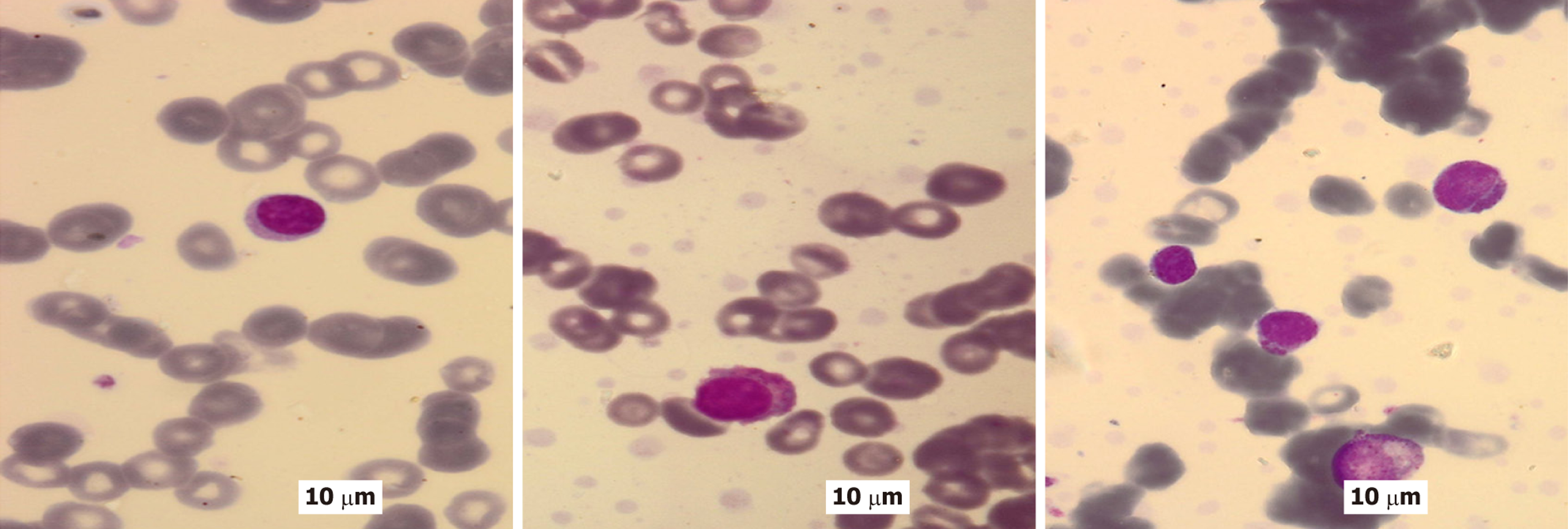
Figure 2 Peripheral blood smears from a patient diagnosed with multiple myeloma with plasma cell leukemia exhibit distinct morph
- Citation: Ebrahim NAA, Elfandy H, Arafat AMA, Darwish AD, Eltohamy MI. Evaluating murine double minute 2 status as a stratification tool for risk-adapted management in plasma cell neoplasms. World J Clin Oncol 2026; 17(1): 111426
- URL: https://www.wjgnet.com/2218-4333/full/v17/i1/111426.htm
- DOI: https://dx.doi.org/10.5306/wjco.v17.i1.111426