
















©The Author(s) 2026.
World J Clin Oncol. Jan 24, 2026; 17(1): 111426
Published online Jan 24, 2026. doi: 10.5306/wjco.v17.i1.111426
Published online Jan 24, 2026. doi: 10.5306/wjco.v17.i1.111426
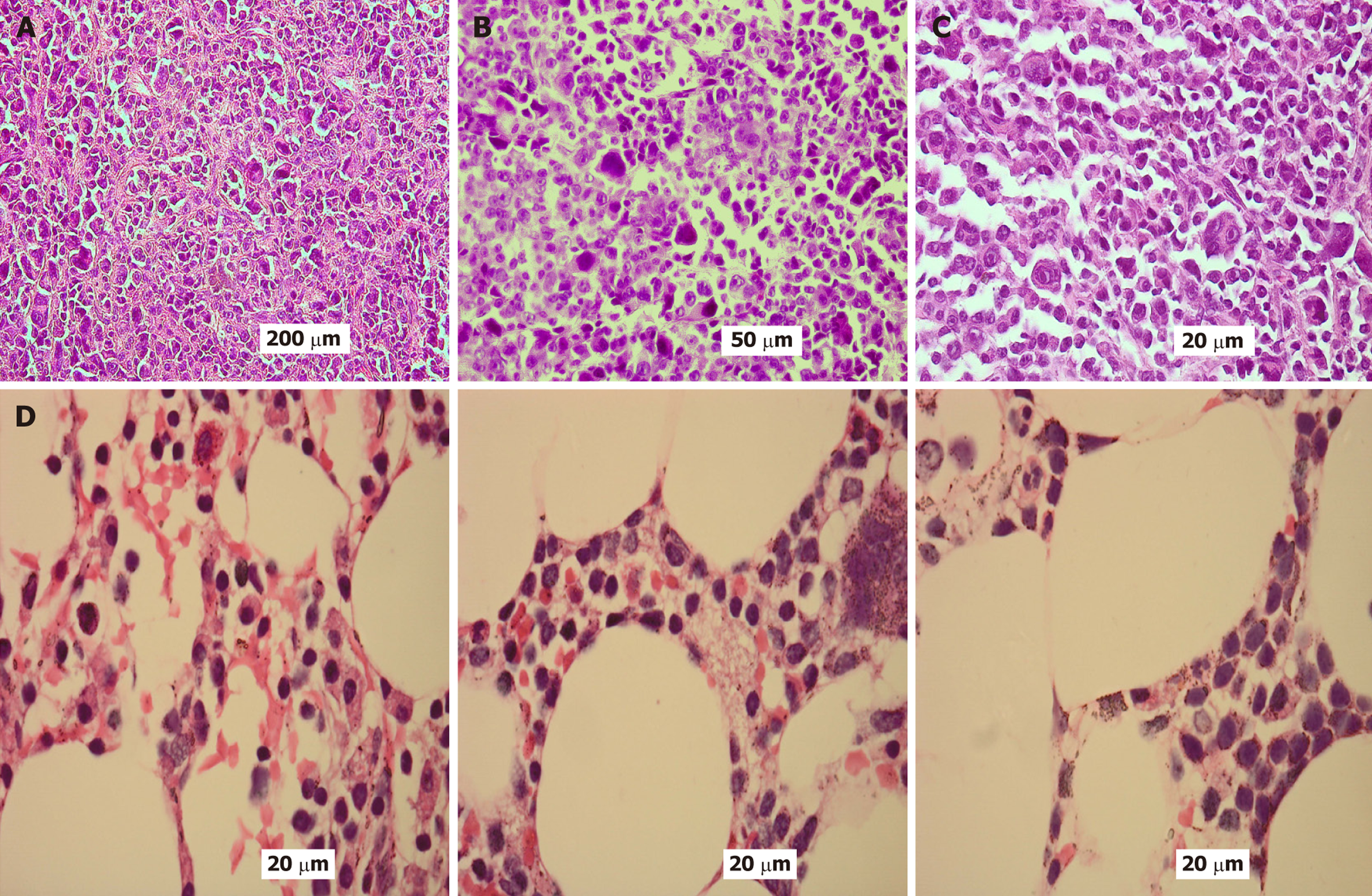
Figure 1 Hematoxylin and eosin.
A-C: Hematoxylin and eosin-stained tissue sections illustrating extramedullary involvement by malignant plasma cells, characteristic of multiple myeloma, shown at progressively increasing magnifications. At low magnification (× 100), the tissue reveals widespread infiltration by densely clustered abnormal plasma cells arranged in continuous sheets, effectively displacing the normal tissue architecture (A). At intermediate magnification (× 200), the neoplastic plasma cells display significant nuclear pleomorphism, frequent binucleation, and scattered mitotic activity, with occasional foci of necrosis observed within the densely cellular environment (B). At high magnification (× 400), individual malignant plasma cells are clearly visualized, showing eccentrically positioned nuclei with a vesicular appearance, prominent nucleoli, distinct perinuclear clearing (Hof), and eosinophilic cytoplasm. Marked nuclear atypia and frequent mitotic figures are evident at this level (C); D: Hematoxylin and eosin-stained bone marrow biopsy sections demonstrate infiltration by myeloma cells in a patient with multiple myeloma with plasma cell leukemia. The marrow notably shows malignant plasma cells that display characteristic morphological features, including eccentrically located nuclei, moderately to abundantly basophilic cytoplasm, and a distinct perinuclear clearing (Hof). Occasional binucleated forms and mitotic figures are observed. Original magnification × 400.
- Citation: Ebrahim NAA, Elfandy H, Arafat AMA, Darwish AD, Eltohamy MI. Evaluating murine double minute 2 status as a stratification tool for risk-adapted management in plasma cell neoplasms. World J Clin Oncol 2026; 17(1): 111426
- URL: https://www.wjgnet.com/2218-4333/full/v17/i1/111426.htm
- DOI: https://dx.doi.org/10.5306/wjco.v17.i1.111426