















©The Author(s) 2025.
World J Clin Oncol. Sep 24, 2025; 16(9): 109644
Published online Sep 24, 2025. doi: 10.5306/wjco.v16.i9.109644
Published online Sep 24, 2025. doi: 10.5306/wjco.v16.i9.109644
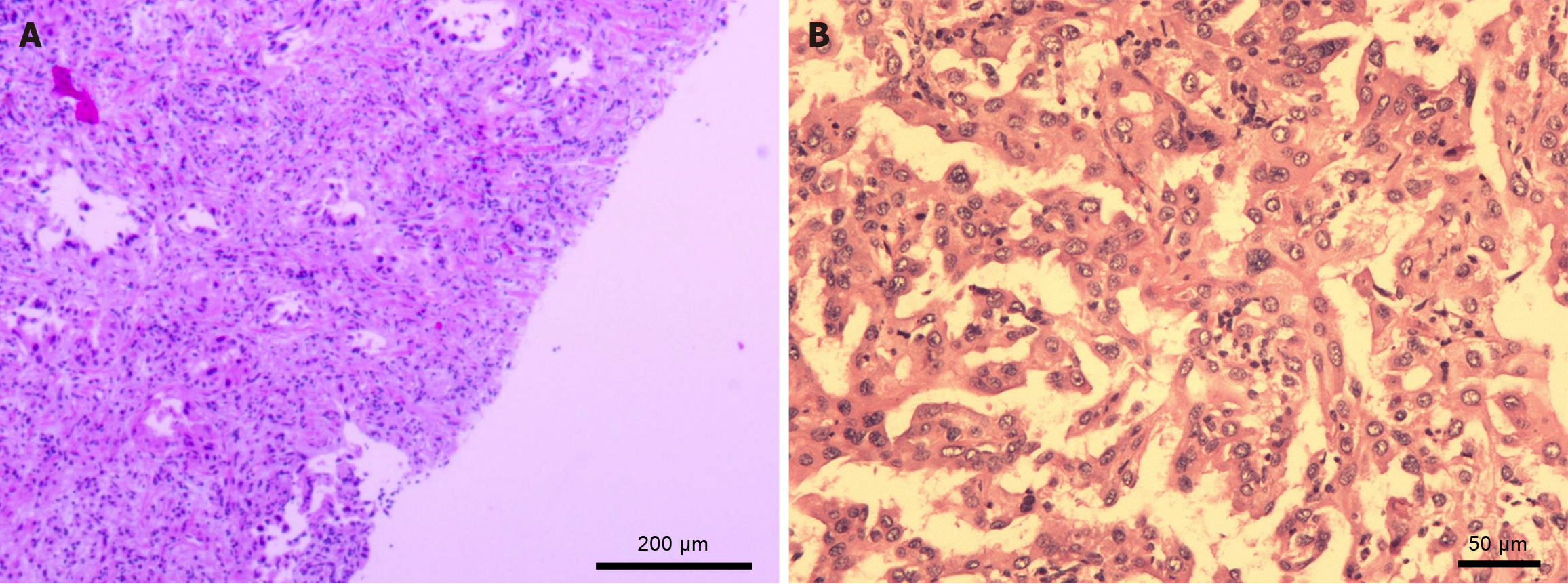
Figure 3 Histopathological confirmation of intrahepatic cholangiocarcinoma.
A: Ultrasound-guided liver biopsy specimen stained with hematoxylin and eosin shows irregular glandular structures with nuclear atypia, which is consistent with adenocarcinoma (scale bar, 200 μm). B: Postoperative resection revealed moderately differentiated intrahepatic cholangiocarcinoma with perineural invasion and dense fibrous stroma (scale bar, 50 μm).
- Citation: Wu ZJ, Wang B, Zhao SC, Pan ZT. Synchronous cholangiocarcinoma and cervical squamous cell carcinoma managed via a multidisciplinary approach: A case report. World J Clin Oncol 2025; 16(9): 109644
- URL: https://www.wjgnet.com/2218-4333/full/v16/i9/109644.htm
- DOI: https://dx.doi.org/10.5306/wjco.v16.i9.109644