















©The Author(s) 2025.
World J Clin Oncol. Sep 24, 2025; 16(9): 109644
Published online Sep 24, 2025. doi: 10.5306/wjco.v16.i9.109644
Published online Sep 24, 2025. doi: 10.5306/wjco.v16.i9.109644
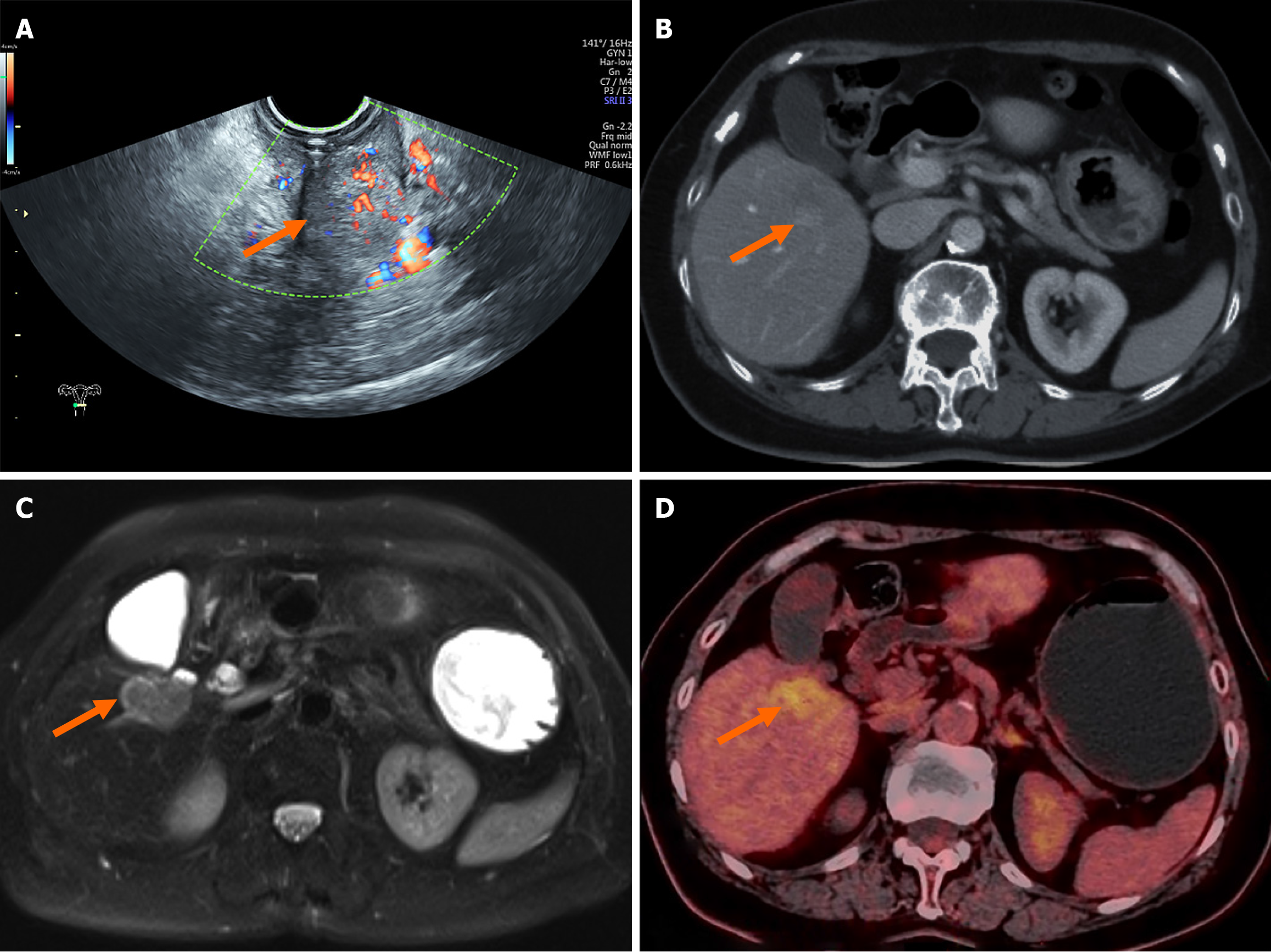
Figure 2 Imaging findings of the cervical and hepatic lesions.
A: Transvaginal ultrasound image showing a hypoechoic cervical mass with poorly defined margins and abundant blood flow (orange arrow); B: Contrast-enhanced computed tomography during the delayed phase showing a hypodense lesion (approximately 3 cm) in segment V of the liver with progressive peripheral enhancement, radiologically consistent with an American Joint Committee on Cancer 8th T1a intrahepatic cholangiocarcinoma (orange arrow); C: Contrast-enhanced magnetic resonance image reveals a hyperintense lesion in segment V on T2-weighted imaging, suggesting malignancy (orange arrow); D: Positron emission tomography/computed tomography image showing intense fluorodeoxyglucose uptake in the same lesion, supporting the diagnosis of a metabolically active primary tumor (orange arrow).
- Citation: Wu ZJ, Wang B, Zhao SC, Pan ZT. Synchronous cholangiocarcinoma and cervical squamous cell carcinoma managed via a multidisciplinary approach: A case report. World J Clin Oncol 2025; 16(9): 109644
- URL: https://www.wjgnet.com/2218-4333/full/v16/i9/109644.htm
- DOI: https://dx.doi.org/10.5306/wjco.v16.i9.109644