Copyright
©The Author(s) 2025.
World J Clin Oncol. Aug 24, 2025; 16(8): 109088
Published online Aug 24, 2025. doi: 10.5306/wjco.v16.i8.109088
Published online Aug 24, 2025. doi: 10.5306/wjco.v16.i8.109088
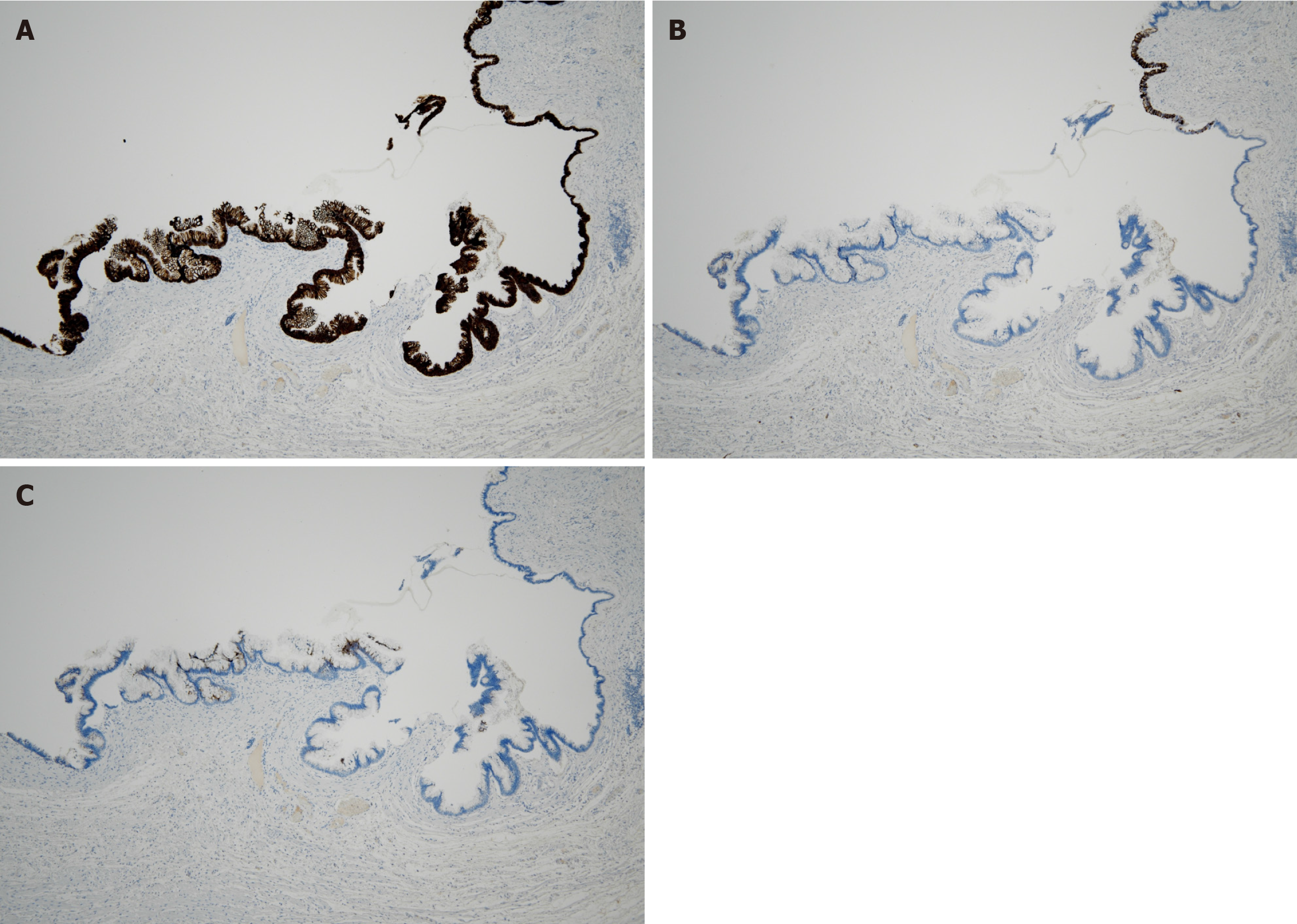
Figure 3 Immunohistochemical findings.
A: Immunohistochemical staining for cytokeratin 20, showing diffuse positivity on the mucosal surface layer (× 40 magnification); B: Immunohistochemical staining of cytokeratin 7, showing partial positivity on the mucosal surface layer (× 40 magnification); C: Immunohistochemical staining for mucin 5AC, showing partial positivity on the mucosal surface layer (× 40 magnification).
- Citation: Mitamura A, Tsujinaka S, Fujishima F, Sawada K, Hikage M, Miura T, Kitamura Y, Hatsuzawa Y, Nakano T, Shibata C. Appendiceal mucinous neoplasms: Optimizing treatment strategies based on clinical, histological, and molecular features. World J Clin Oncol 2025; 16(8): 109088
- URL: https://www.wjgnet.com/2218-4333/full/v16/i8/109088.htm
- DOI: https://dx.doi.org/10.5306/wjco.v16.i8.109088


