
















©The Author(s) 2025.
World J Clin Oncol. Aug 24, 2025; 16(8): 109088
Published online Aug 24, 2025. doi: 10.5306/wjco.v16.i8.109088
Published online Aug 24, 2025. doi: 10.5306/wjco.v16.i8.109088
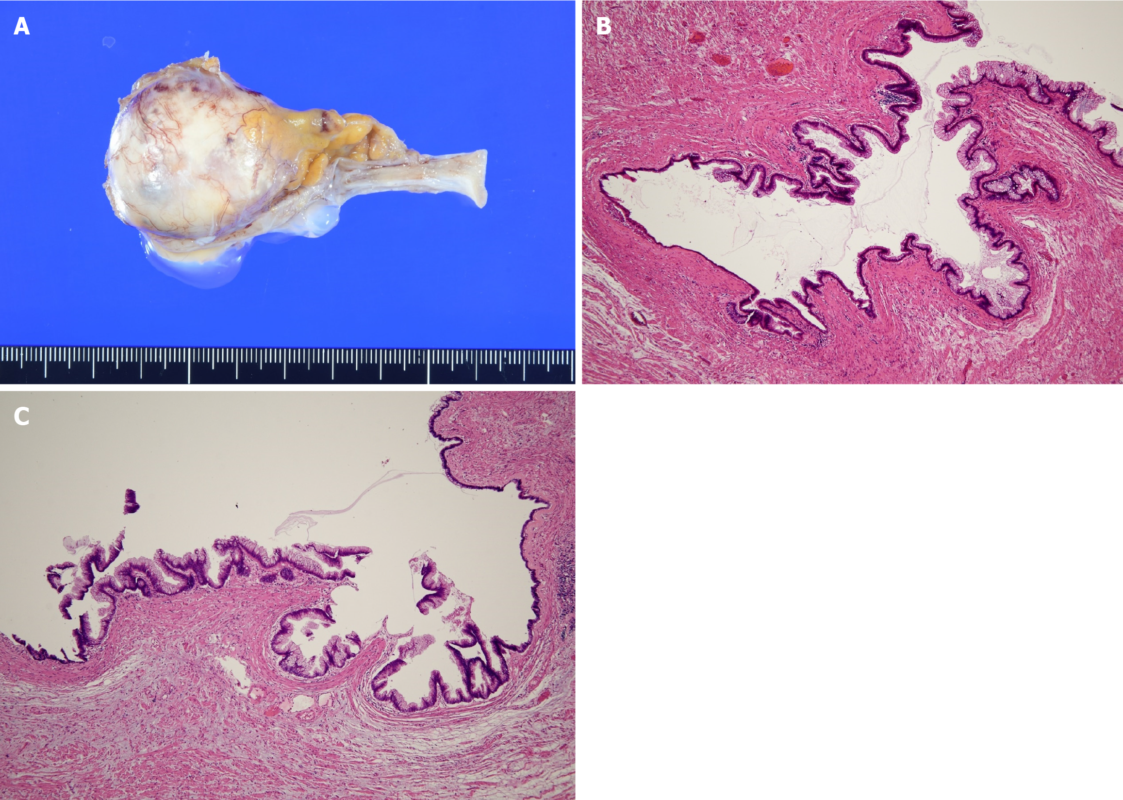
Figure 2 Macroscopic and microscopic findings.
A: Macroscopic view of a low-grade appendiceal mucinous neoplasm (LAMN), showing cystic dilation of the appendix filled with mucinous content; B: Microscopic view of a LAMN with hematoxylin & eosin staining (× 40 magnification), showing a mucosal surface lined by a single layer of mucin-producing columnar epithelium without high-grade cytologic atypia; C: Additional microscopic view of a LAMN with hematoxylin & eosin staining (× 40 magnification), highlighting features similar to panel B.
- Citation: Mitamura A, Tsujinaka S, Fujishima F, Sawada K, Hikage M, Miura T, Kitamura Y, Hatsuzawa Y, Nakano T, Shibata C. Appendiceal mucinous neoplasms: Optimizing treatment strategies based on clinical, histological, and molecular features. World J Clin Oncol 2025; 16(8): 109088
- URL: https://www.wjgnet.com/2218-4333/full/v16/i8/109088.htm
- DOI: https://dx.doi.org/10.5306/wjco.v16.i8.109088