
















©The Author(s) 2025.
World J Clin Oncol. Aug 24, 2025; 16(8): 107208
Published online Aug 24, 2025. doi: 10.5306/wjco.v16.i8.107208
Published online Aug 24, 2025. doi: 10.5306/wjco.v16.i8.107208
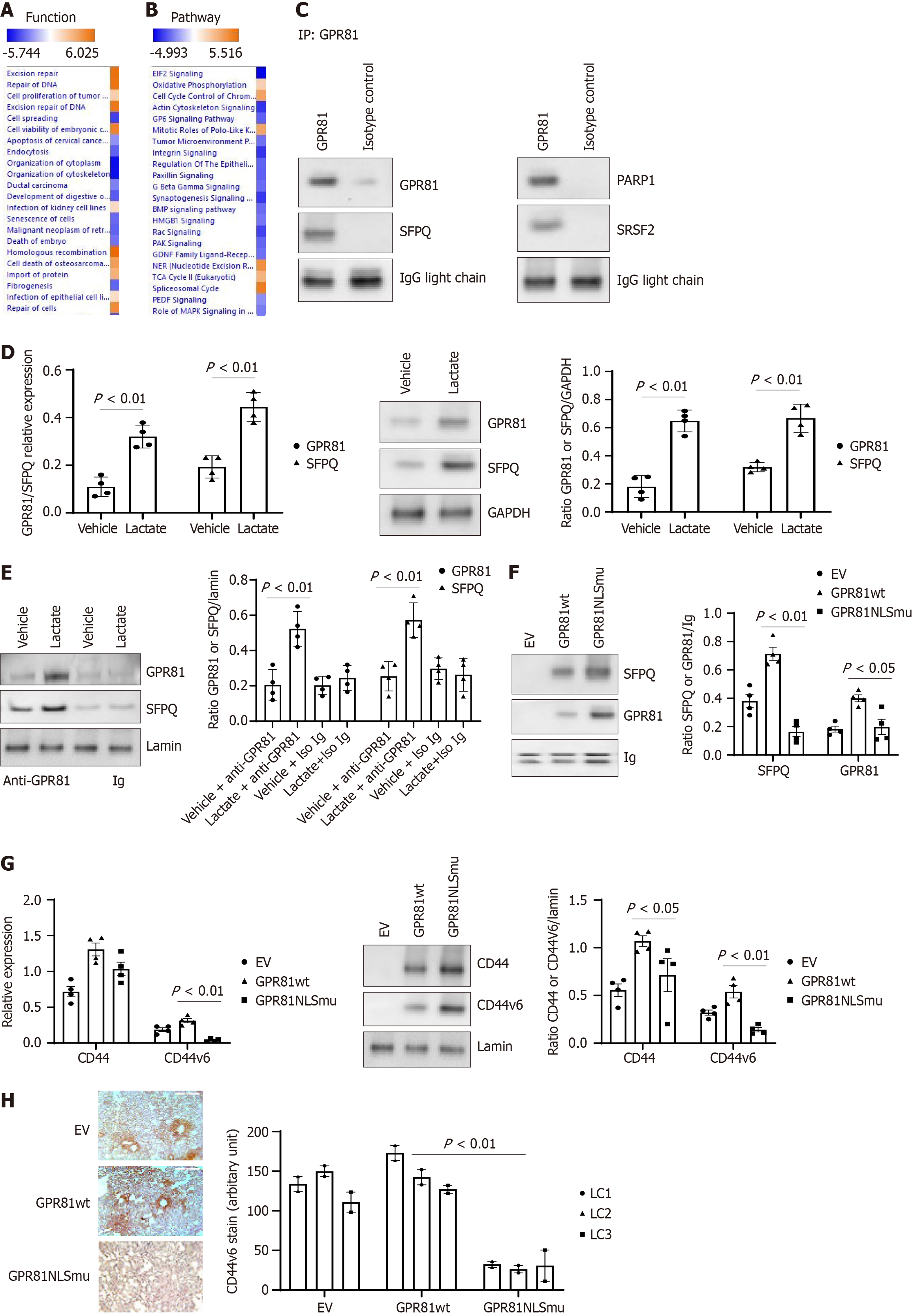
Figure 5 The interaction of GPR81 with other nuclear proteins affects lung cancer cell function.
Proteins identified from GPR81 immunoprecipitated proteins in lung cancer cells in Proteomics analysis were applied to Ingenuity Pathway Analysis (IPA). A: Top functions associated with the lung cancer cell dataset as shown by IPA pathway analysis; B: Top canonical pathways associated with proteins from the lung cancer cell dataset as shown by IPA pathway analysis. Cell functions, or pathways identified are represented on the y-axis. The x-axis corresponds to the –log of the P-value (Fisher’s exact test) and the orange points on each pathway bar represent the ratio of the number of proteins in a given pathway that meet the cutoff criteria, divided by the total number of proteins that map to that pathway; C: Immunoprecipitation and western blot analysis with GPR81 bound proteins. The nuclear fraction from A549 was used in immunoprecipitation with the GPR81 antibody. Then SFPQ, PARP1 and SRSF2 antibodies were used in western blot analysis with the GPR81 pull down portion of the lung cancer nuclear fraction. Lactate promotes SFPQ expression and formation of GPR81/SFPQ complex in lung cancer cells; D: GPR81 reverse transcriptase-PCR (RT-PCR) was conducted with lung cancer cells cultured under vehicle/Lactate (5 mmol/L) conditions. (left panel). GPR81 antibody was used in immunoprecipitation with lung cancer cells cultured under vehicle/Lactate (5 mmol/L) conditions and the SFPQ levels were measured with western blot (middle panel). Densitometry values summarizing western blot data are shown in the right panel. GAPDH served as a loading control; E: GPR81 immunoprecipitation was conducted with the cell nuclear fraction from lung cancer cell cultured under vehicle/Lactate (5 mmol/L) conditions. Then the protein bands were detected with anti-GPR81 and SFPQ antibodies. Western blot was shown in the left panel. Densitometry values summarizing western blot data are shown in the right panel. Lamin served as a loading control; F: Lung cancer cells transduced with Lenti virus with empty vector, GPR81 wild-type or GPR81 NLS mutant, antibody was used in immunoprecipitation with lung cancer cell nuclear fraction; G: The same set of cells was used to analyze CD44 and CD44v6 expression. The CD44 and CD44v6 Levels were measured with RT-PCR (left panel) and western blot (middle panel). Densitometry values summarizing western blot data are shown in the right panel. GAPDH served as a loading control. Lung cancer cell A549, H661, H858, H645 were used in figure D-G; H: Anti-CD44v6 was used to assess the distribution of human cells expressing CD44v6 protein expressing cells from mice lung regions from mice receiving lung cancer cells transduced with empty vector, GPR81 wild-type (GPR81wt) (middle panel) or GPR81 NLS mutant (GPR81NLSmu) (right panel). Scale bar = 200 μmol/L. Quantification was summed up in the bottom figure. Image J was used to quantify CD44v6 protein positive cells in lung cancer cell explanted mice lung tissue sections.
- Citation: Yang L, Kono T, Gilbertsen A, Li Y, Sun B, Jacobson BA, Karam S, Dehm SM, Henke CA, Kratzke RA. GPR81 nuclear transportation is critical for cancer growth and progression in lung and other solid cancers. World J Clin Oncol 2025; 16(8): 107208
- URL: https://www.wjgnet.com/2218-4333/full/v16/i8/107208.htm
- DOI: https://dx.doi.org/10.5306/wjco.v16.i8.107208