Copyright
©The Author(s) 2025.
World J Clin Oncol. Aug 24, 2025; 16(8): 107208
Published online Aug 24, 2025. doi: 10.5306/wjco.v16.i8.107208
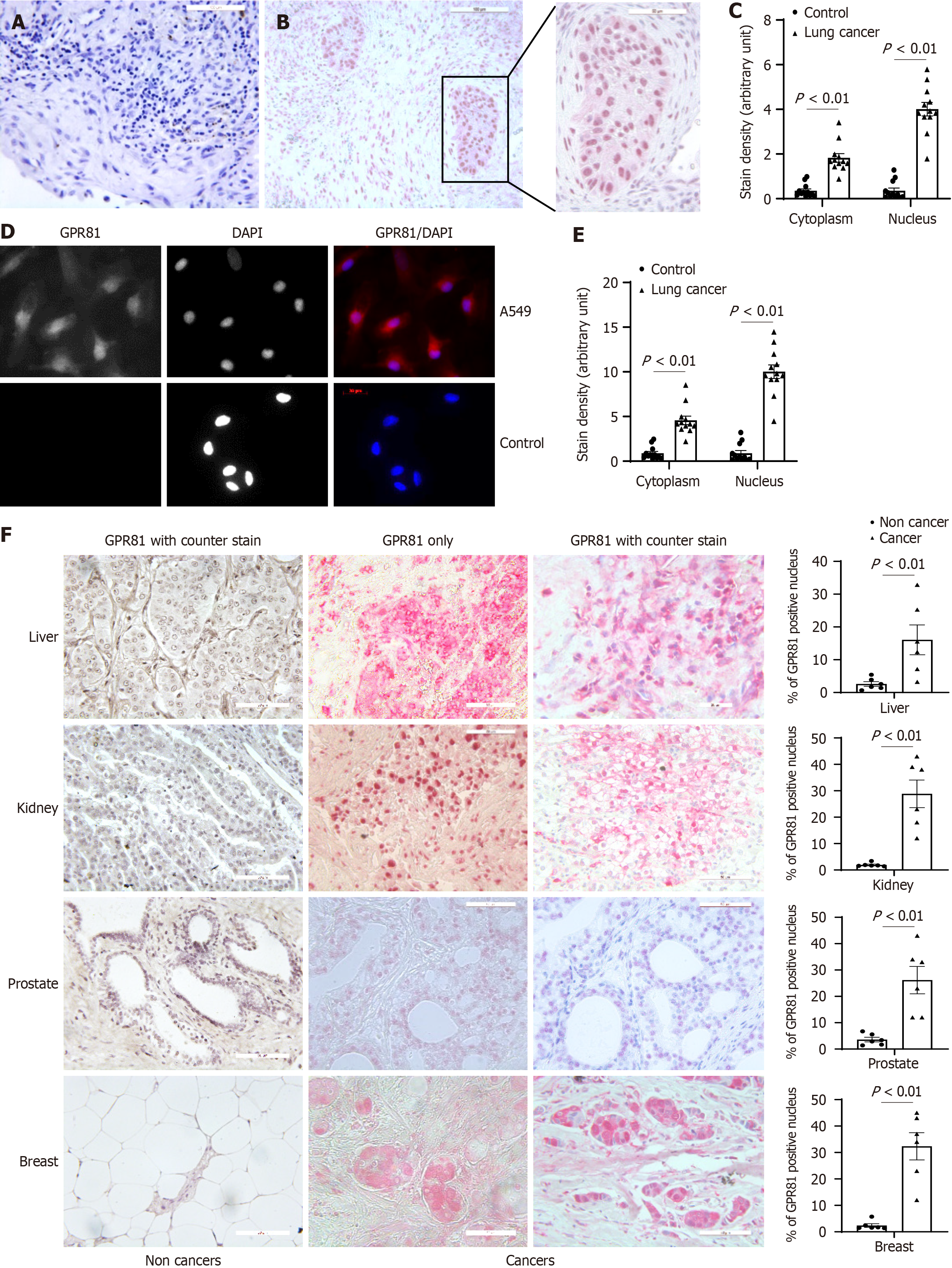
Figure 2 Nuclear GPR81 is present in non-small cell lung cancer cells and in non-small cell lung cancer tissue sections.
Immuno histochemistry (IHC) stain was performed on human lung tissue. A and B: Control and lung cancer. Tissue sections were used in IHC immunostain. IHC was performed using antibodies to GPR81 to display GPR81 distribution in control and cancer tissue sections (red); Counterstain was Mayer’s hematoxylin to display the nucleus (blue). Scale bar = 100 µm; C: GPR81 stain was quantified with Image J and summarized in figure C. Twelve each of control and non-small cell lung cancer (NSCLC) patient specimens, 5 sections were imaged in each specimen; D: Immunoflurescent stain was performed on human NSCLC and control cells with GPR81 antibody. Scale bar = 20 μmol/L; E: GPR81 stain in figure D was quantified with Image J and summarized (4 cell lines each, 202, 205, 272, 392, A549, H661, H858, H645); F: Tissue sections were used in IHC immunostain. IHC was performed using antibodies to GPR81 to display GPR81 distribution in control and cancer tissue section (red); left panels are example images for non cancer liver, prostate, kidney and breast tissue; the middle two panels are example images for liver cancer, prostate cancer, kidney cancer and breast cancer (scale bar = 100 μmol/L). The right panel is a summary of the IHC stain for those cancers. Counterstain was Mayer’s hematoxylin to display the nucleus (blue). Scale bar = 50 µm.
- Citation: Yang L, Kono T, Gilbertsen A, Li Y, Sun B, Jacobson BA, Karam S, Dehm SM, Henke CA, Kratzke RA. GPR81 nuclear transportation is critical for cancer growth and progression in lung and other solid cancers. World J Clin Oncol 2025; 16(8): 107208
- URL: https://www.wjgnet.com/2218-4333/full/v16/i8/107208.htm
- DOI: https://dx.doi.org/10.5306/wjco.v16.i8.107208