Copyright
©The Author(s) 2025.
World J Clin Oncol. Jul 24, 2025; 16(7): 109246
Published online Jul 24, 2025. doi: 10.5306/wjco.v16.i7.109246
Published online Jul 24, 2025. doi: 10.5306/wjco.v16.i7.109246
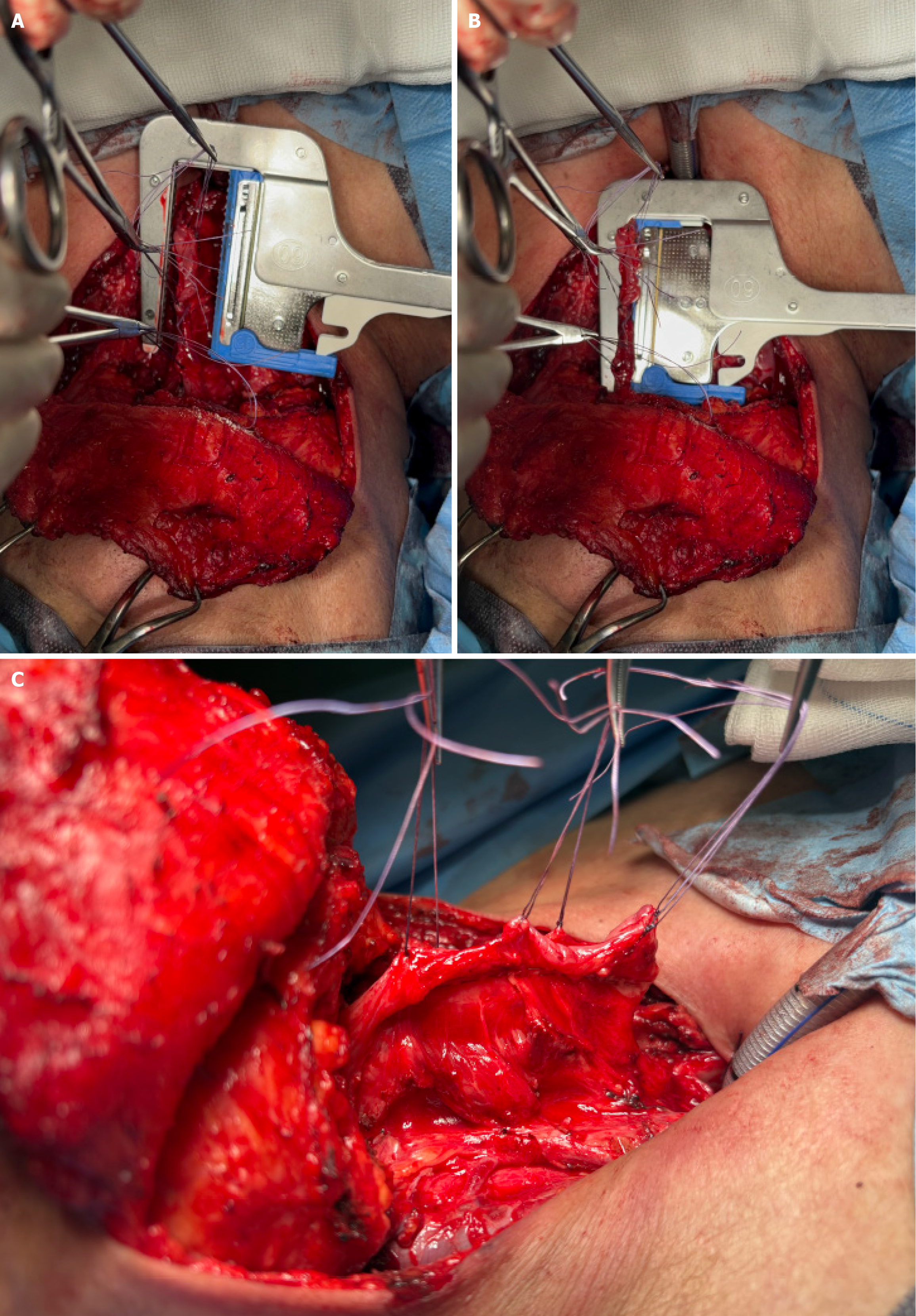
Figure 3 Intraoperative images demonstrating the application of a surgical stapler following total laryngectomy.
Intraoperative images illustrating the application of a surgical stapler. A and B: The stapler is positioned on the esophageal mucosa (A), which is then (B) clamped to ensure precise closure; C: The final result shows two parallel rows of staples securing the sutured pharyngeal mucosa, indicating a well-aligned closure.
- Citation: Galazka A, Stawarz K, Bienkowska-Pluta K, Paszkowska M, Misiak-Galazka M. Closure techniques for esophageal reconstruction after total laryngectomy and their impact on fistula formation. World J Clin Oncol 2025; 16(7): 109246
- URL: https://www.wjgnet.com/2218-4333/full/v16/i7/109246.htm
- DOI: https://dx.doi.org/10.5306/wjco.v16.i7.109246


