Copyright
©The Author(s) 2025.
World J Clin Oncol. Jul 24, 2025; 16(7): 104663
Published online Jul 24, 2025. doi: 10.5306/wjco.v16.i7.104663
Published online Jul 24, 2025. doi: 10.5306/wjco.v16.i7.104663
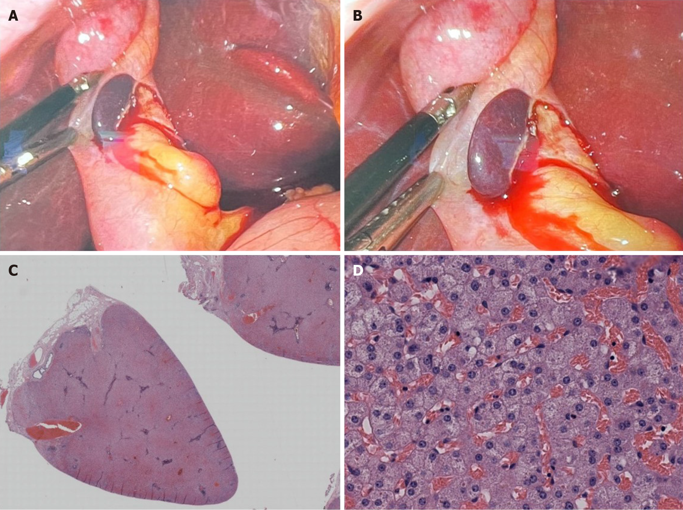
Figure 4 Intra-operative and high-power microscopic image of ectopic liver tissue.
A-D: Intra-operative images of ectopic liver tissue during cholecystectomy (A and B) and high-power microscopic image of ectopic liver tissue with sickle cell congestion (C and D, Hematoxylin & eosin, 40 ×).
- Citation: Saikia K, Xu Z, Azordegan N, Ahsan BU. Incidental diagnosis of gallbladder carcinoma during or after routine cholecystectomy: A retrospective study with emphasis on clinicopathologic findings. World J Clin Oncol 2025; 16(7): 104663
- URL: https://www.wjgnet.com/2218-4333/full/v16/i7/104663.htm
- DOI: https://dx.doi.org/10.5306/wjco.v16.i7.104663


