Copyright
©The Author(s) 2025.
World J Clin Oncol. May 24, 2025; 16(5): 104762
Published online May 24, 2025. doi: 10.5306/wjco.v16.i5.104762
Published online May 24, 2025. doi: 10.5306/wjco.v16.i5.104762
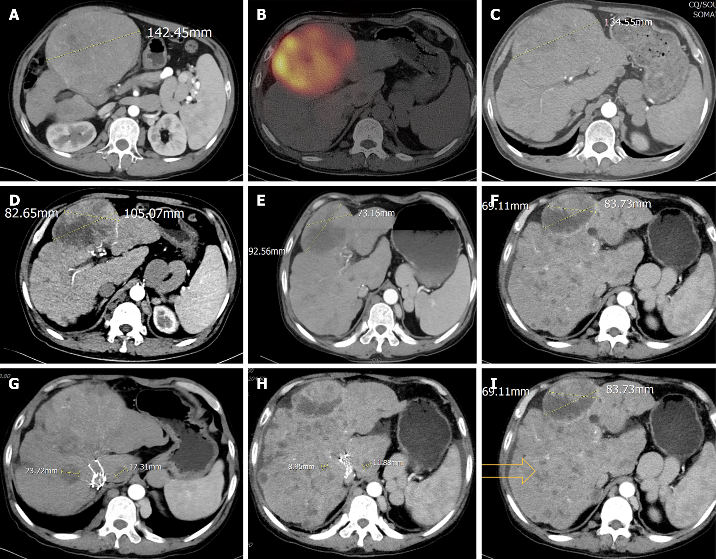
Figure 1 Imaging characteristics of the patient.
A: Enhanced computed tomography showed a 142.45 mm exophytic hepatocellular carcinoma located at left medial lobe of the liver with multiple intrahepatic metastases at initial diagnosis; B: The distribution of yttrium-90 microspheres within tumors; C: After a single session of hepatic arterial infusion chemotherapy, the tumor size measured 134.55 mm, which decreased a little; D: Four months post-treatment, the target tumor size declined to 105.07 mm progressively. Visible areas of necrosis increased. The maximum diameter of the viable lesion reduced to 82.65 mm; E: Six months post-treatment, the target tumor shrank notably with the maximum diameter of the viable tumor measuring 7316 mm, gradually growing necrosis areas, but little enlargement of non-target intrahepatic lesions; F: Ten months post-treatment, the target lesion continues to shrink in size, and the liver volume has increased. However, multiple new nodular lesions have emerged within the liver. Figures 1A-F primarily illustrate the changes in size and tumor viability of the target lesion; G and H: The diameter changes of measurable non-target lesions before treatment and at the last follow-up visit, indicating significant reductions; I: The hollow arrows indicate the multiple small nodular lesions that newly appeared within the liver at the time of the last computed tomography scan.
- Citation: Shao MH, Tan BB, Chen HL, Zhang H. Yttrium-90 radioembolization for advanced hepatocellular carcinoma with Budd-Chiari syndrome: A case report. World J Clin Oncol 2025; 16(5): 104762
- URL: https://www.wjgnet.com/2218-4333/full/v16/i5/104762.htm
- DOI: https://dx.doi.org/10.5306/wjco.v16.i5.104762


