Copyright
©The Author(s) 2025.
World J Clin Oncol. Apr 24, 2025; 16(4): 105077
Published online Apr 24, 2025. doi: 10.5306/wjco.v16.i4.105077
Published online Apr 24, 2025. doi: 10.5306/wjco.v16.i4.105077
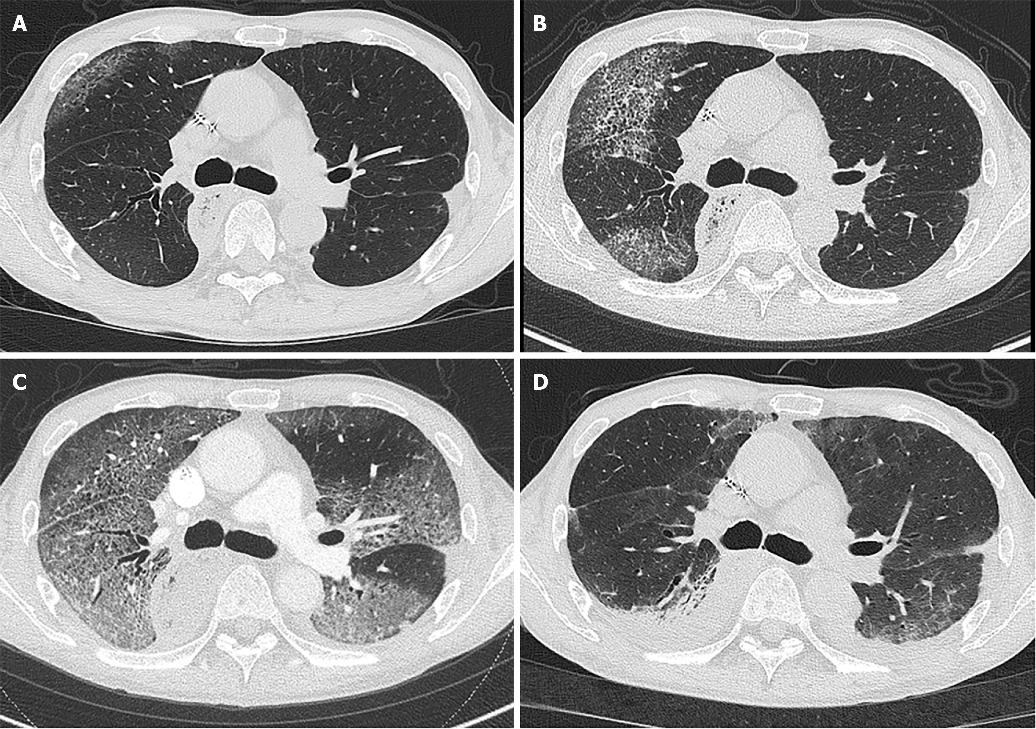
Figure 1 Computed tomography scan of the lungs.
A: At the initial presentation. Ground glass opacity was seen in a limited area of both lungs; B: On admission. The ground glass opacity expanded and involved about 20% of the total lung volume; C: On day 3 of admission. Almost all lung fields are opacified; D: On day 12 of admission. The opacities are dramatically improved.
- Citation: Tsurui T, Mura E, Horiike A, Tsunoda T. Oxaliplatin-induced diffuse alveolar hemorrhage: A case report. World J Clin Oncol 2025; 16(4): 105077
- URL: https://www.wjgnet.com/2218-4333/full/v16/i4/105077.htm
- DOI: https://dx.doi.org/10.5306/wjco.v16.i4.105077


