
















©The Author(s) 2025.
World J Clin Oncol. Mar 24, 2025; 16(3): 102301
Published online Mar 24, 2025. doi: 10.5306/wjco.v16.i3.102301
Published online Mar 24, 2025. doi: 10.5306/wjco.v16.i3.102301
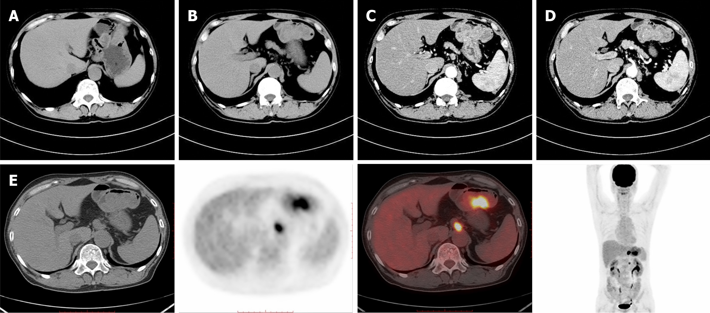
Figure 2 Pre-operation abdominal computed tomography images and positron emission tomography-computed tomography.
A and B: Abdominal computed tomography (CT) revealed the gastric wall at the greater curvature of remnant stomach were obviously thickened; C and D: Enhanced CT revealed the gastric wall at the greater curvature of remnant stomach were obviously thickened; E: Positron emission tomography-CT showed metabolism nodular thickening of the residual gastric wall, enlarged lymph nodes beside the abdominal aorta (at the T12 level) and in the middle abdomen (at the L2 level).
- Citation: Wang T, Cheng Y, Hu F, Wang Q. Residual gastric cancer with a mixed small cell neuroendocrine and keratinizing squamous cell carcinoma: A case report. World J Clin Oncol 2025; 16(3): 102301
- URL: https://www.wjgnet.com/2218-4333/full/v16/i3/102301.htm
- DOI: https://dx.doi.org/10.5306/wjco.v16.i3.102301