
















©The Author(s) 2025.
World J Clin Oncol. Dec 24, 2025; 16(12): 111701
Published online Dec 24, 2025. doi: 10.5306/wjco.v16.i12.111701
Published online Dec 24, 2025. doi: 10.5306/wjco.v16.i12.111701
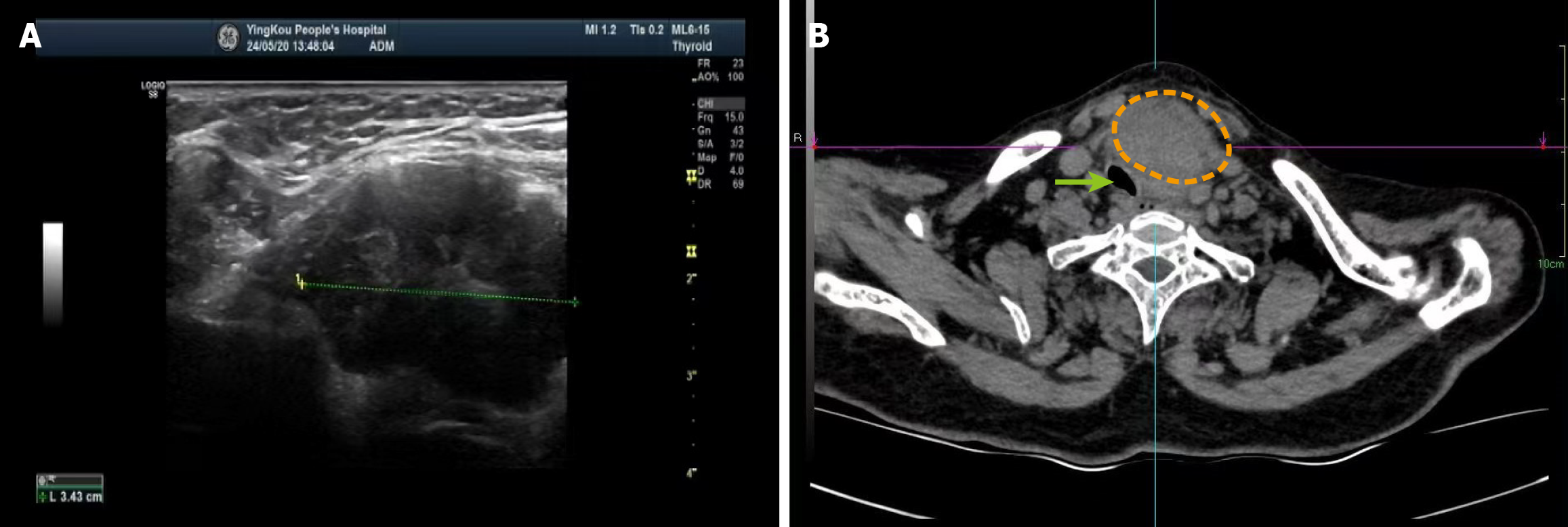
Figure 4 Imaging examinations.
A: Thyroid ultrasonography showing a mass with regular morphology and well-defined margins, closely associated with the lower pole of the thyroid gland; B: Contrast-enhanced axial computed tomography scan of the neck demonstrating a large mass causing tracheal deviation. The orange dotted line outlines the approximate tumor extent, and the green arrow indicates the deviated trachea.
- Citation: Shan BW, Yu JH, Ren T. Intrathyroidal thymic carcinoma comprising squamous cell and small cell carcinoma components: A case report. World J Clin Oncol 2025; 16(12): 111701
- URL: https://www.wjgnet.com/2218-4333/full/v16/i12/111701.htm
- DOI: https://dx.doi.org/10.5306/wjco.v16.i12.111701