
















©The Author(s) 2025.
World J Clin Oncol. Dec 24, 2025; 16(12): 111701
Published online Dec 24, 2025. doi: 10.5306/wjco.v16.i12.111701
Published online Dec 24, 2025. doi: 10.5306/wjco.v16.i12.111701
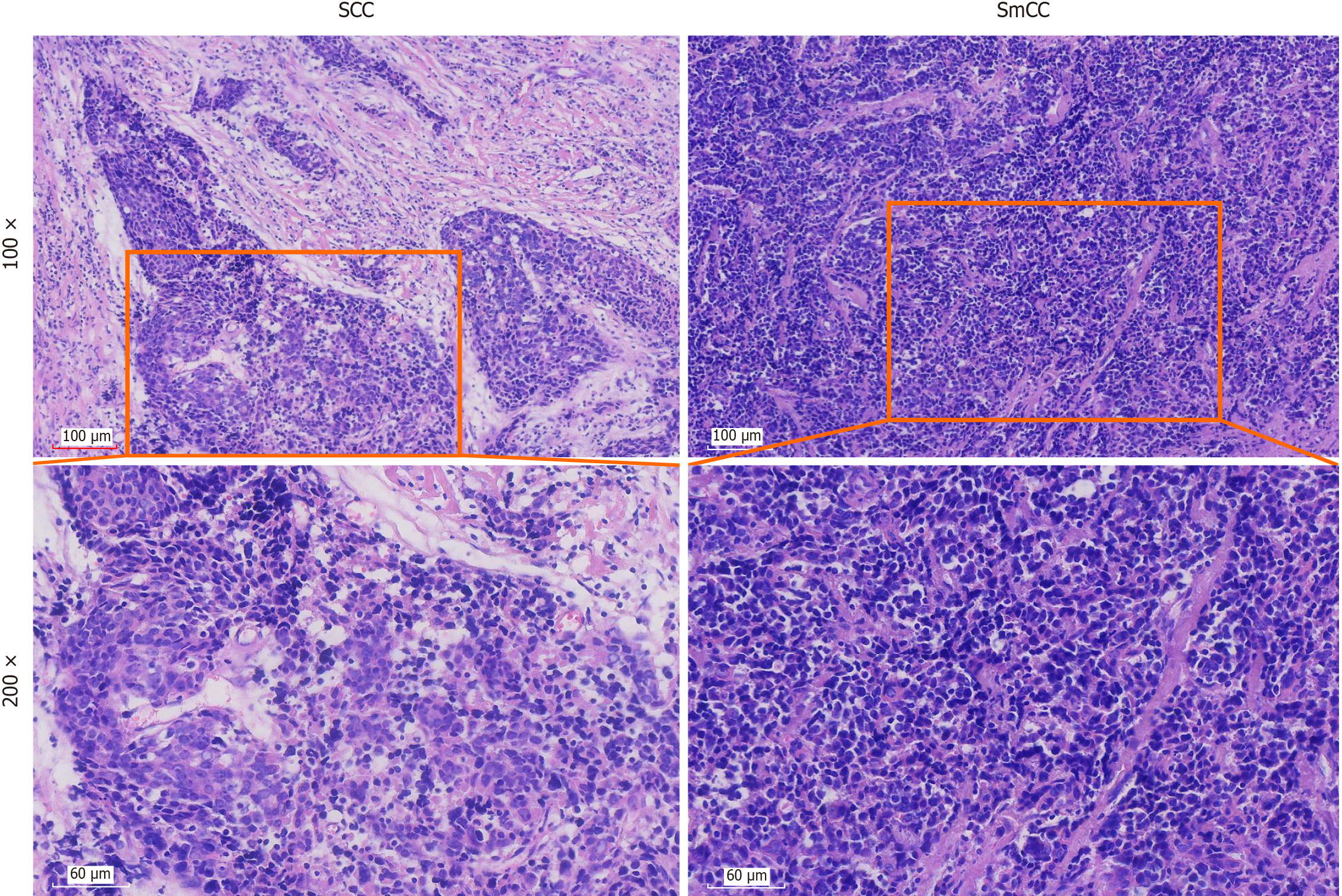
Figure 1 The histomorphology of the tumour reveals two distinct components: Squamous cell carcinoma and small cell carcinoma.
In the squamous cell carcinoma region: At 100 × magnification, abundant stroma components are visible, with nest of tumour cells forming islands separated by the stroma; at 200 × magnification, the tumour cells exhibit slightly eosinophilic cytoplasm, pale vacuolated nuclei, and prominent nucleoli. In the small cell carcinoma region: At 100 × magnification, the tumour cells are arranged in sheets, with minimal surrounding stroma; at 200 × magnification, the tumour cells display scanty cytoplasm, poorly defined cell borders, finely granular and lightly stained chromatin, and inconspicuous nucleoli. Scale bar is 60 μm. SCC: Squamous cell carcinoma; SmCC: Small cell carcinoma.
- Citation: Shan BW, Yu JH, Ren T. Intrathyroidal thymic carcinoma comprising squamous cell and small cell carcinoma components: A case report. World J Clin Oncol 2025; 16(12): 111701
- URL: https://www.wjgnet.com/2218-4333/full/v16/i12/111701.htm
- DOI: https://dx.doi.org/10.5306/wjco.v16.i12.111701