
















©The Author(s) 2025.
World J Clin Oncol. Nov 24, 2025; 16(11): 111764
Published online Nov 24, 2025. doi: 10.5306/wjco.v16.i11.111764
Published online Nov 24, 2025. doi: 10.5306/wjco.v16.i11.111764
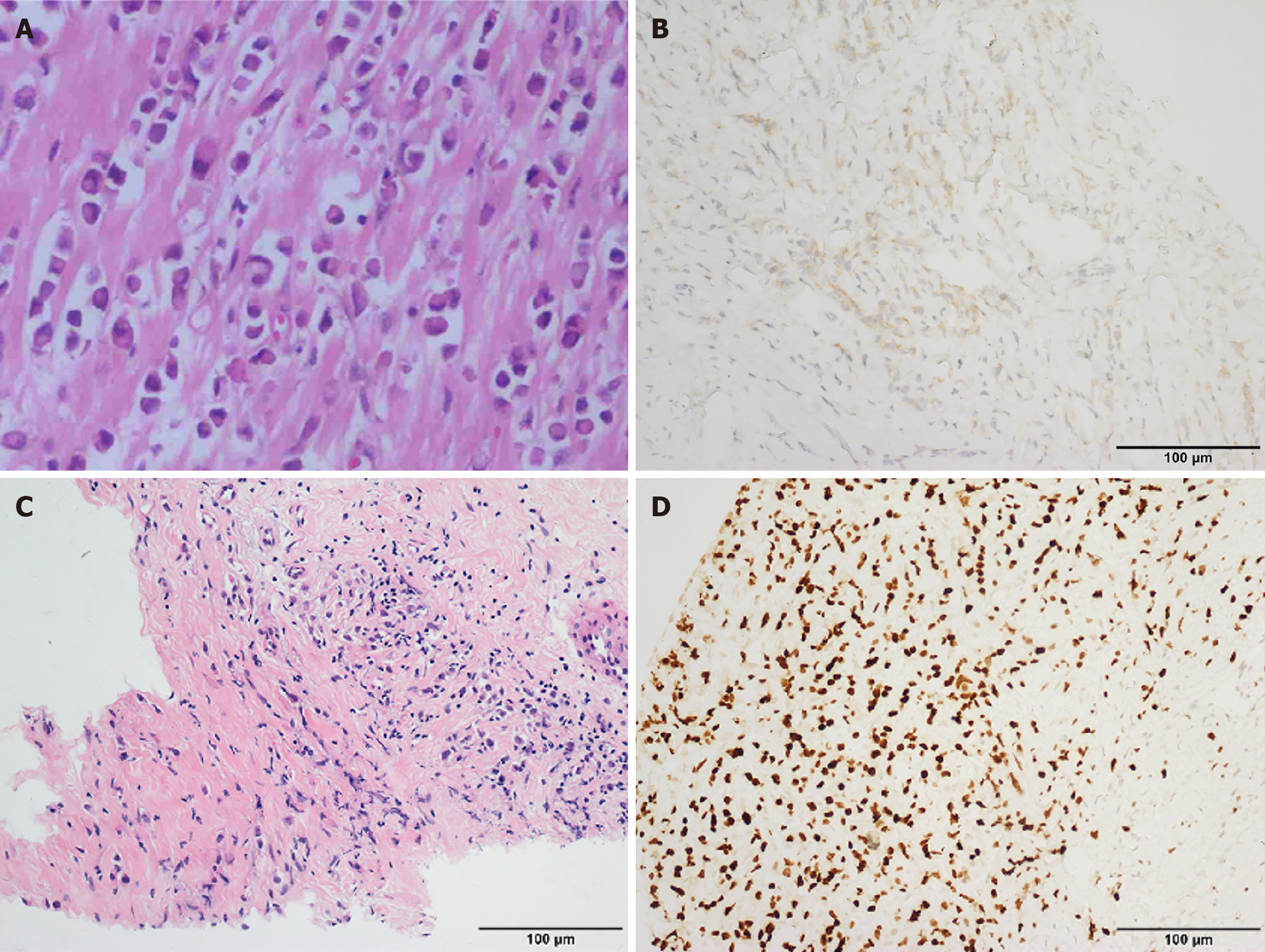
Figure 4 Pathological findings.
A: Hematoxylin and eosin (HE) staining at 200 × magnification revealed invasive lobular carcinoma of the breast, with visible lymphovascular tumor emboli and neural invasion; B: Immunohistochemical staining for HER2 in lymph nodes. Out of 23 Lymph nodes examined, cancer was present in 22, HER2(+); C: Cardia puncture tissue pathological HE staining at 200 × magnification: In the fibrous tissue, tumor is observed, which has the same morphological characteristics as the tumor found in the breast tissue section that was resected and examined in January 2016; D: Immunohistochemical staining for GATA3, confirming the metastatic origin of the tumor from the breast. The findings are consistent with the metastatic spread of lobular carcinoma of the breast.
- Citation: Pan HY, Liu W, Ding W, Wang ZM, Feng YY, Yu AH, Cheng CS. Dynamic esophageal manometry reveals pseudoachalasia secondary to metastatic breast cancer: A case report. World J Clin Oncol 2025; 16(11): 111764
- URL: https://www.wjgnet.com/2218-4333/full/v16/i11/111764.htm
- DOI: https://dx.doi.org/10.5306/wjco.v16.i11.111764