
















©The Author(s) 2025.
World J Clin Oncol. Nov 24, 2025; 16(11): 111527
Published online Nov 24, 2025. doi: 10.5306/wjco.v16.i11.111527
Published online Nov 24, 2025. doi: 10.5306/wjco.v16.i11.111527
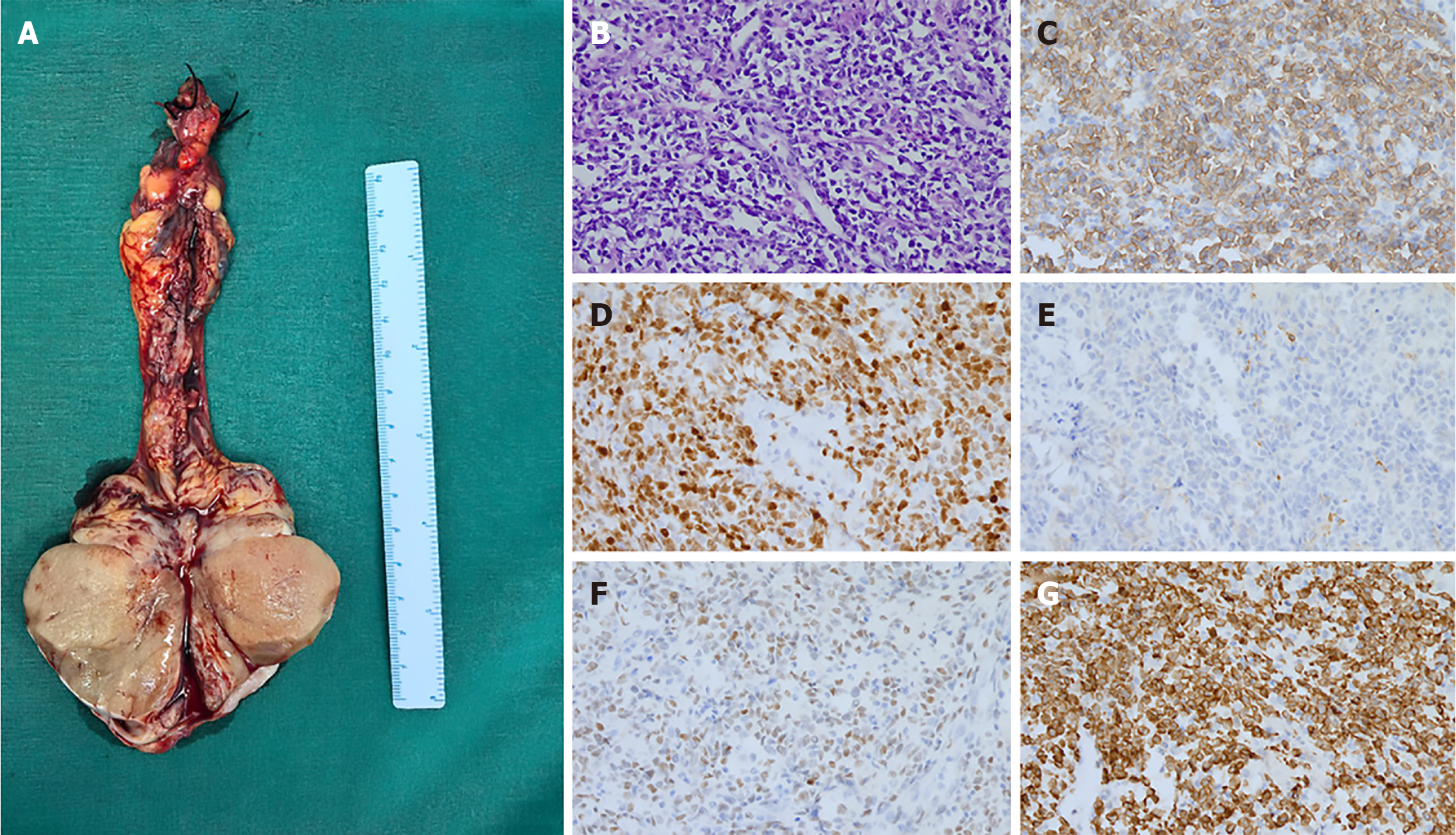
Figure 2 Pathological findings of left testis tumor.
A: Orchiectomy specimen showing an enlarged testis and spermatic cord tumor; B: Lymphatic cellular infiltration with monomorphic, medium-to-large round cells and nuclear pleomorphism, H&E (× 40). C-G: Immunohistochemistry findings. C: MUM1 (× 20, weak positive); D: CD10 (× 20, negative); E: BCL6 (× 20, negative); F: CD20 (× 20, strong positive); G: Ki-67 (× 20, approximately 80% positive, high proliferation rate).
- Citation: Zuo YZ, Liang Z, Pan BJ, Yan WG, Zhou ZE. Primary testicular diffuse large B-cell lymphoma with gonadal vein tumor thrombus: A case report and review of the literature. World J Clin Oncol 2025; 16(11): 111527
- URL: https://www.wjgnet.com/2218-4333/full/v16/i11/111527.htm
- DOI: https://dx.doi.org/10.5306/wjco.v16.i11.111527