
















©The Author(s) 2025.
World J Clin Oncol. Nov 24, 2025; 16(11): 111527
Published online Nov 24, 2025. doi: 10.5306/wjco.v16.i11.111527
Published online Nov 24, 2025. doi: 10.5306/wjco.v16.i11.111527
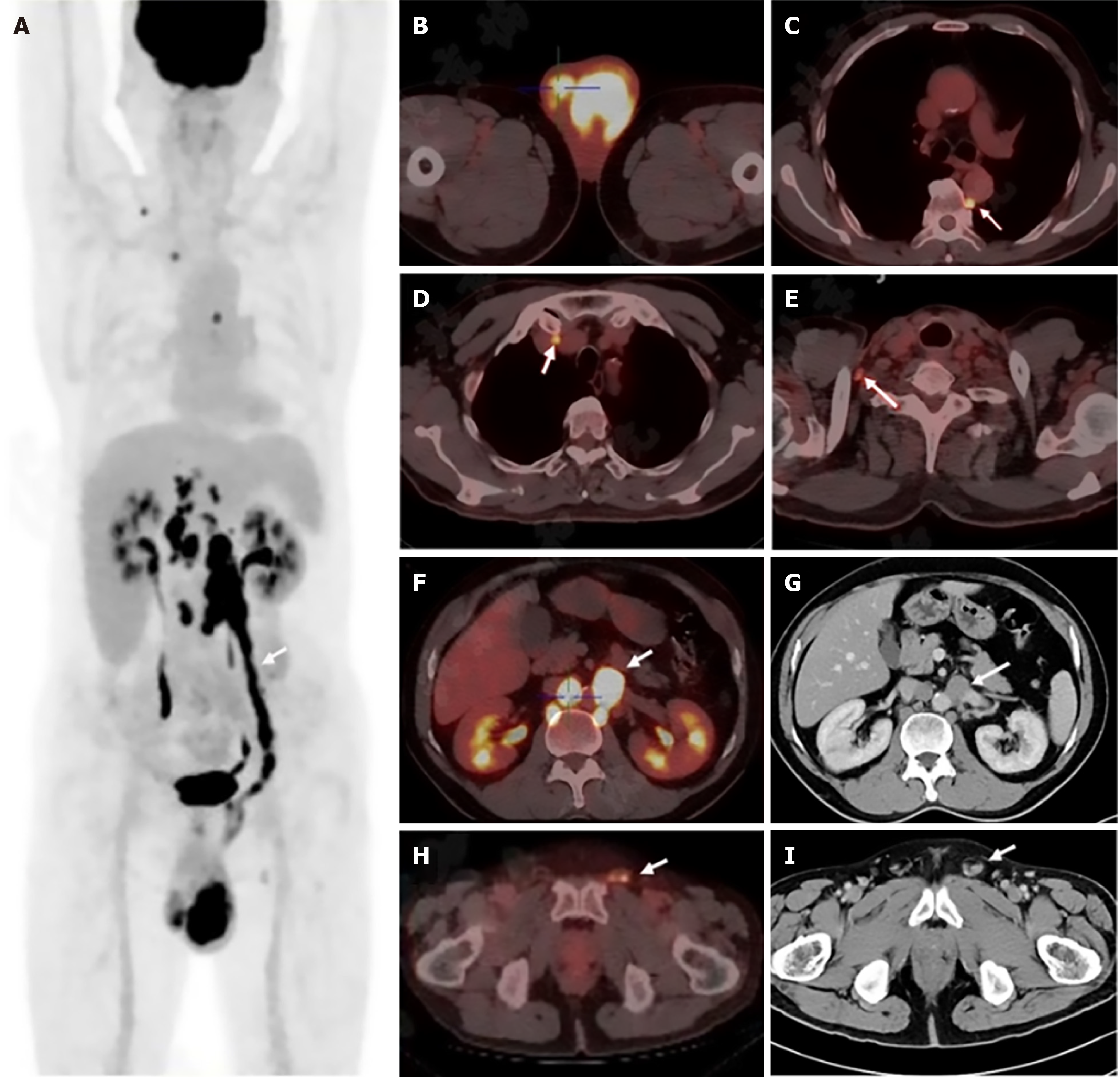
Figure 1 18F-fluorodeoxyglucose positron emission tomography/computed tomography and contrast-enhanced computed tomography images.
A: Linear uptake along the left gonadal vein (arrow, SUVmax 16.5); B: Bilateral testicular lesions (left: SUVmax 14.1; right: SUVmax 5.0); C: Paravertebral lymph node uptake (arrow); D: Mediastinal lymph node uptake (arrow); E: Right supraclavicular lymph node uptake (arrow); F and G: Tumor thrombus in the left gonadal vein near the left renal vein (arrow); H and I: Tumor thrombus in the left spermatic cord (arrow).
- Citation: Zuo YZ, Liang Z, Pan BJ, Yan WG, Zhou ZE. Primary testicular diffuse large B-cell lymphoma with gonadal vein tumor thrombus: A case report and review of the literature. World J Clin Oncol 2025; 16(11): 111527
- URL: https://www.wjgnet.com/2218-4333/full/v16/i11/111527.htm
- DOI: https://dx.doi.org/10.5306/wjco.v16.i11.111527