
















©The Author(s) 2025.
World J Clin Oncol. Nov 24, 2025; 16(11): 110257
Published online Nov 24, 2025. doi: 10.5306/wjco.v16.i11.110257
Published online Nov 24, 2025. doi: 10.5306/wjco.v16.i11.110257
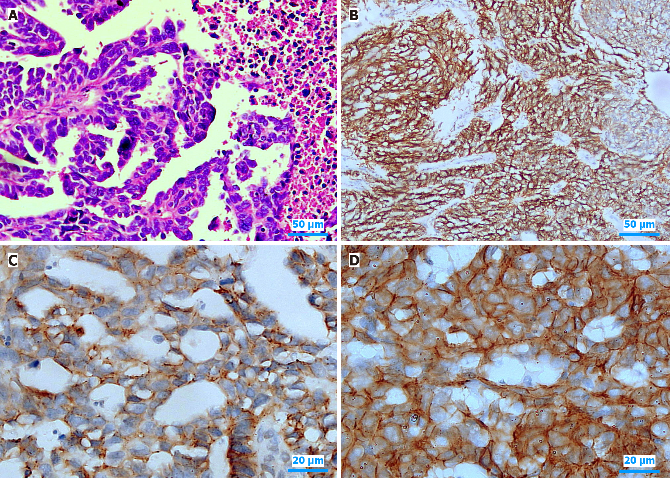
Figure 1 Representative histological and immunohistochemical images depicting claudin-6 expression in high-grade endometrial serous carcinoma.
A: Hematoxylin and eosin (H&E)-stained section (magnification × 400) demonstrating a complex arrangement of papillary and micropapillary structures lined by highly atypical epithelial cells. These tumor cells display pronounced nuclear pleomorphism, hyperchromasia, conspicuous nucleoli, and frequent mitotic figures, which are classic histopathological features of high-grade serous carcinoma; B: Low-power immunohistochemical view (DAB chromogen, × 100) illustrating diffuse, intense membranous Claudin-6 staining distributed throughout clusters of malignant epithelial cells; C: Higher magnification field (× 400) revealing heterogeneous membranous claudin-6 expression, with areas showing incomplete and variable staining intensity along the tumor cell membrane; D: Another high-power field (× 400) demonstrating consistently strong, continuous circumferential membranous staining for Claudin-6 in densely arranged neoplastic cells. Scale bar = 50 μm for A and B, 20 μm for C and D.
- Citation: Ebrahim NAA, Eissa TS, Hussein MA, Korany OM, Amin NH. Tight junction disruption via claudin-6 overexpression promotes invasion and recurrence in high-grade endometrial tumors. World J Clin Oncol 2025; 16(11): 110257
- URL: https://www.wjgnet.com/2218-4333/full/v16/i11/110257.htm
- DOI: https://dx.doi.org/10.5306/wjco.v16.i11.110257