
















©The Author(s) 2025.
World J Clin Oncol. Nov 24, 2025; 16(11): 110202
Published online Nov 24, 2025. doi: 10.5306/wjco.v16.i11.110202
Published online Nov 24, 2025. doi: 10.5306/wjco.v16.i11.110202
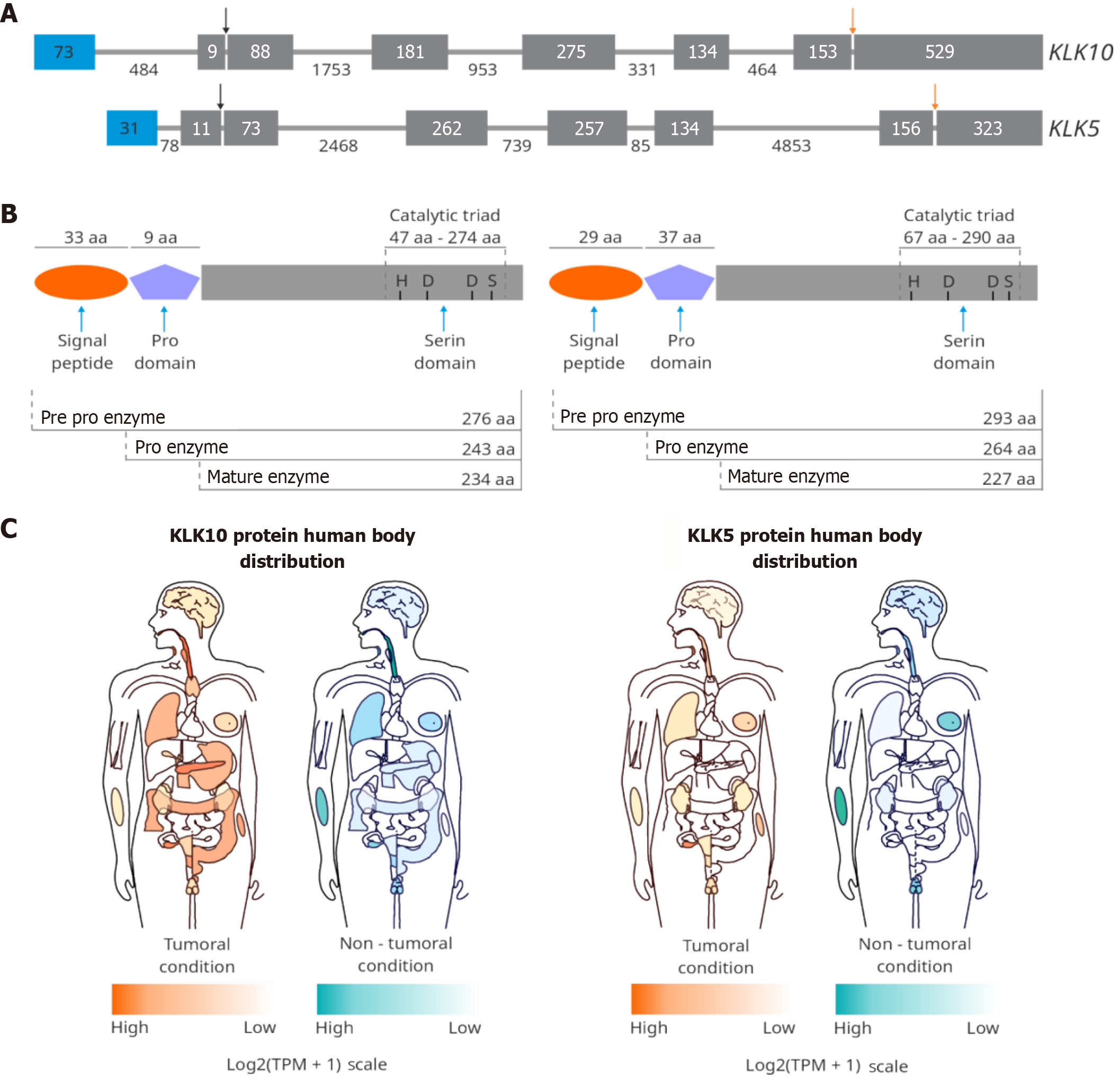
Figure 2 Schematic representation of kallikrein-related peptidases 10 and 5 and distribution in the human body in normal and cancer conditions[93].
The blue color represents healthy conditions, and the orange color represents tumor conditions. A higher intensity of color correlated with a higher expression of kallikrein-related peptidases (KLK) 10 or KLK5 in the indicated organ. A: KLK10 and KLK5 gene structure. Both genes have five coding exons (gray boxes) and one non-coding exon (blue boxes). The length (in base pairs) of exons and introns is indicated inside and outside the boxes, respectively. The black row represents the position of the start codon, and the orange row indicates the stop codon’s position; B: Protein structure of KLK10 and KLK5; C: Schema modified of GEPIA web server for the expression of KLK10 and KLK5 in the human body. Citation: Tang Z, Li C, Kang B, Gao G, Li C, Zhang Z. GEPIA: A web server for cancer and normal gene expression profiling and interactive analyses. Nucleic Acids Res 2017; 45: W98-W102. Copyright ©The Authors 2025. Published by Oxford University Press. (https://academic.oup.com/pages/open-research/open-access). KLK: Kallikrein-related peptidases; TPM: Transcripts per million.
- Citation: Palacios Serrato EG, Medina-Abreu KH, Oropeza-Martínez E, Jacinto-Alemán LF, Macías-Silva M, Tecalco-Cruz AC. ADAMTS-8 and kallikrein-related peptidases 10 and 5 proteases also have a tumor suppression role. World J Clin Oncol 2025; 16(11): 110202
- URL: https://www.wjgnet.com/2218-4333/full/v16/i11/110202.htm
- DOI: https://dx.doi.org/10.5306/wjco.v16.i11.110202