
















©The Author(s) 2025.
World J Clin Oncol. Oct 24, 2025; 16(10): 108876
Published online Oct 24, 2025. doi: 10.5306/wjco.v16.i10.108876
Published online Oct 24, 2025. doi: 10.5306/wjco.v16.i10.108876
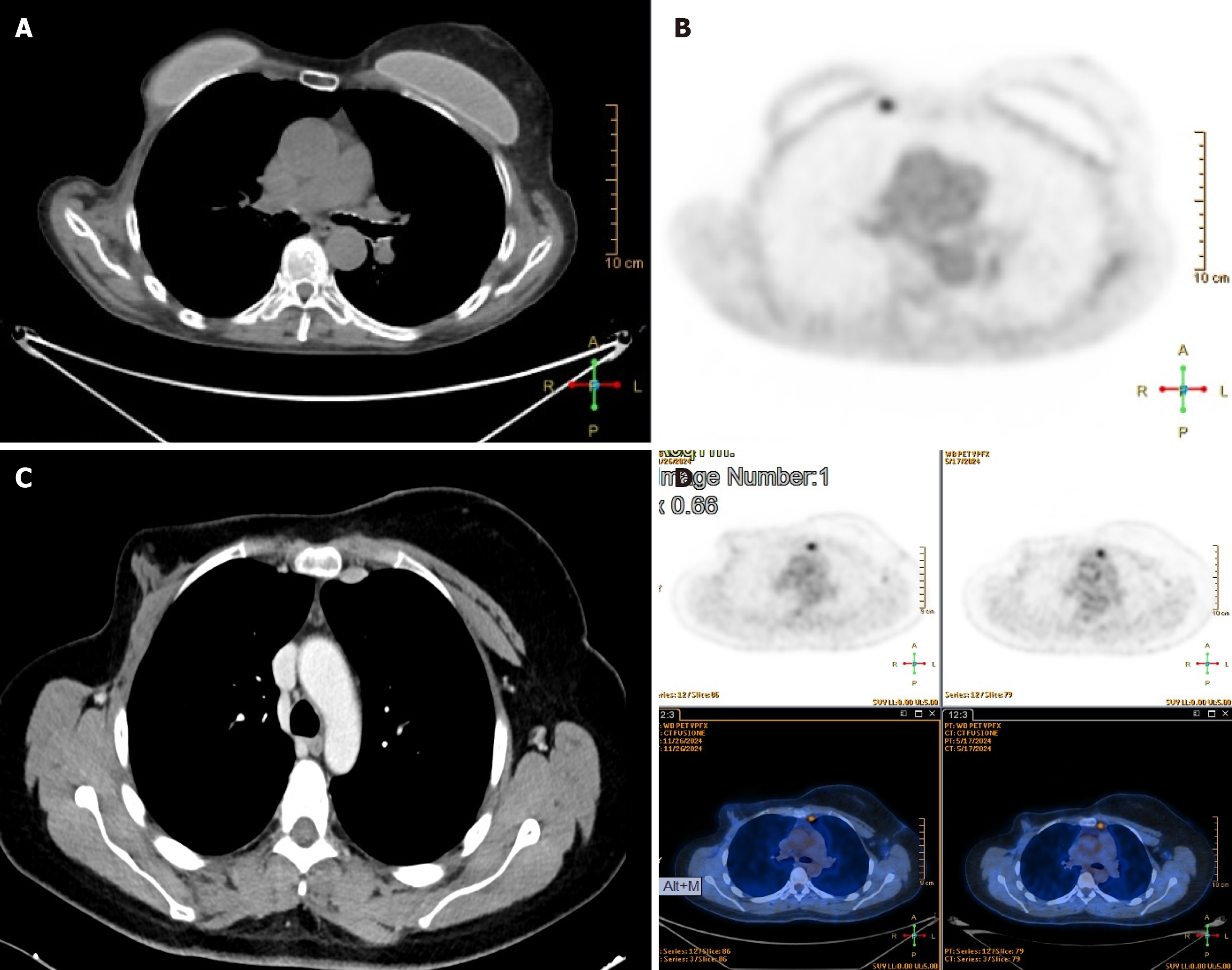
Figure 2 Chest computed tomography scan and positron emission tomography scan.
A and B: Left internal mammary lymphadenopathy; C and D: Right internal mammary lymphadenopathy.
- Citation: Pardolesi A, Ferrari M, Leuzzi G, Stanzi A, Calderoni M, Uslenghi C, Scarci M, Raveglia F, Cioffi U, Solli P. Robotic approach for lymphadenectomy of internal mammary lymph nodes in breast cancer: Five case reports. World J Clin Oncol 2025; 16(10): 108876
- URL: https://www.wjgnet.com/2218-4333/full/v16/i10/108876.htm
- DOI: https://dx.doi.org/10.5306/wjco.v16.i10.108876