Copyright
©The Author(s) 2022.
World J Clin Oncol. Jun 24, 2022; 13(6): 540-552
Published online Jun 24, 2022. doi: 10.5306/wjco.v13.i6.540
Published online Jun 24, 2022. doi: 10.5306/wjco.v13.i6.540
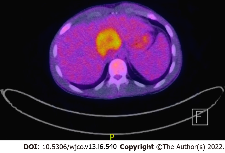
Figure 4 Positron emission tomography scan showing a hypermetabolic mass arising from the medial segment of the left liver lobe, measuring about 5.
1 cm x 4.7 cm in the axial and anteroposterior dimension and 6.9 cm in the craniocaudal dimension in case 2.
- Citation: Khan AA, Estfan BN, Yalamanchali A, Niang D, Savage EC, Fulmer CG, Gosnell HL, Modaresi Esfeh J. Epstein-Barr virus-associated smooth muscle tumors in immunocompromised patients: Six case reports. World J Clin Oncol 2022; 13(6): 540-552
- URL: https://www.wjgnet.com/2218-4333/full/v13/i6/540.htm
- DOI: https://dx.doi.org/10.5306/wjco.v13.i6.540


