Copyright
©The Author(s) 2021.
World J Clin Oncol. Dec 24, 2021; 12(12): 1227-1243
Published online Dec 24, 2021. doi: 10.5306/wjco.v12.i12.1227
Published online Dec 24, 2021. doi: 10.5306/wjco.v12.i12.1227
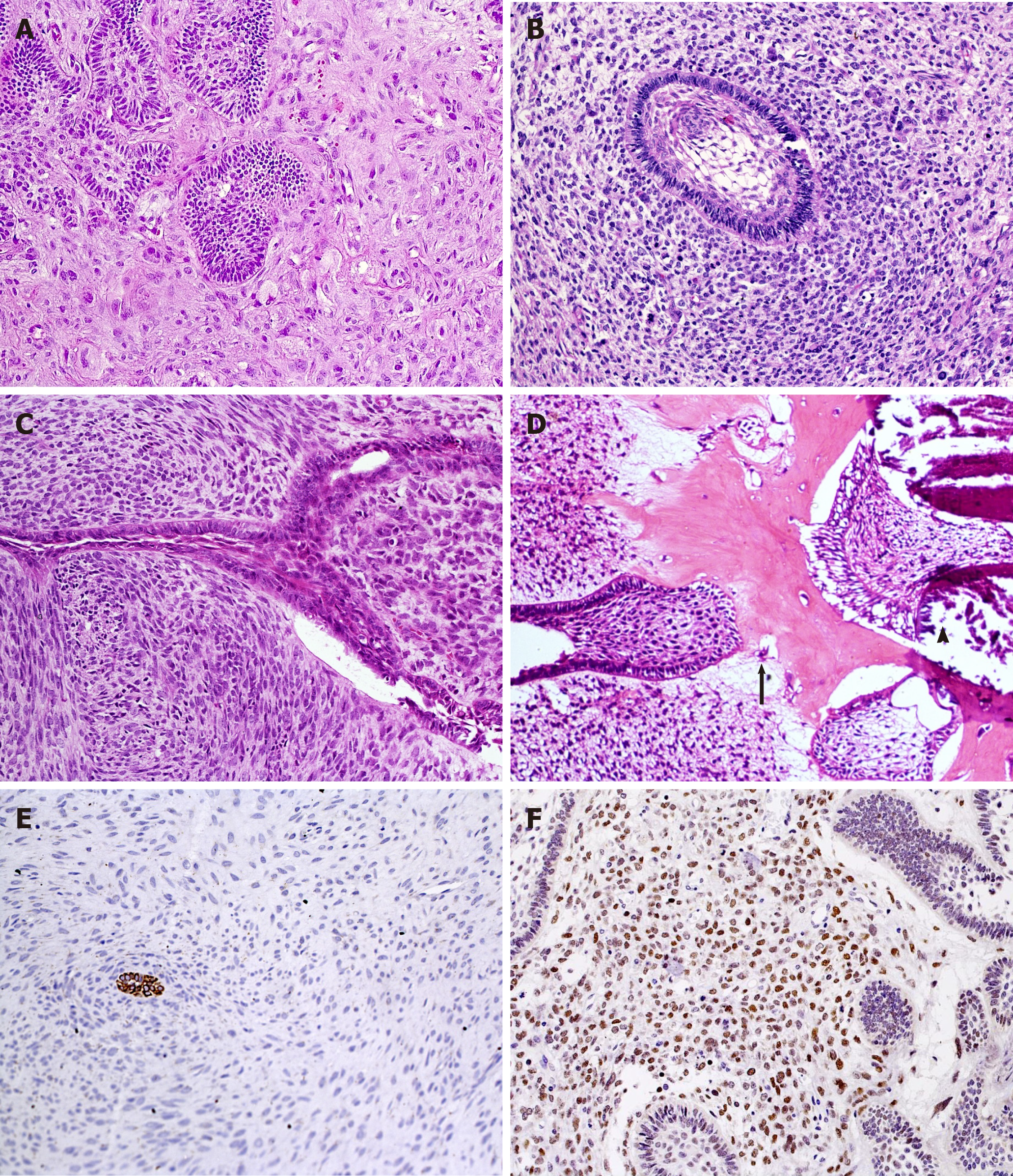
Figure 3 Histopathological aspects of ameloblastic fibromas and ameloblastic fibro-odontosarcoma.
Marked pleomorphism and atypia in mesenchymal cells (HE, 20×) (A-C). A: Several mitotic figures, nuclear hyperchromatism and multinucleated and aberrant cells are seen in a highly pleomorphic sarcomatous component of ameloblastic fibromas, while epithelial islands remain benign; B: A follicular benign epithelial island is surrounded by hypercellularized sarcomatous proliferation; C: Malignant mesenchymal tissue resembling a storiform pattern and haphazard disposition of sarcomatous cells around an epithelial branching cord; D: Production of enamel matrix (arrowhead) and dentinoid (arrow) as well as malignant mesenchymal tissue (left side) are components of this ameloblastic fibro-odontosarcoma (A-D: HE, 20×); E: Cytokeratins can help to localize odontogenic epithelial cells within dominant sarcomatous proliferation (IHC for AE1/AE3, 20×); F: Most mesenchymal malignant cells show nuclear positivity for p53 antigen (IHC for p53, 20×).
- Citation: Sánchez-Romero C, Paes de Almeida O, Bologna-Molina R. Mixed odontogenic tumors: A review of the clinicopathological and molecular features and changes in the WHO classification. World J Clin Oncol 2021; 12(12): 1227-1243
- URL: https://www.wjgnet.com/2218-4333/full/v12/i12/1227.htm
- DOI: https://dx.doi.org/10.5306/wjco.v12.i12.1227


