Copyright
©The Author(s) 2021.
World J Clin Oncol. Oct 24, 2021; 12(10): 960-965
Published online Oct 24, 2021. doi: 10.5306/wjco.v12.i10.960
Published online Oct 24, 2021. doi: 10.5306/wjco.v12.i10.960
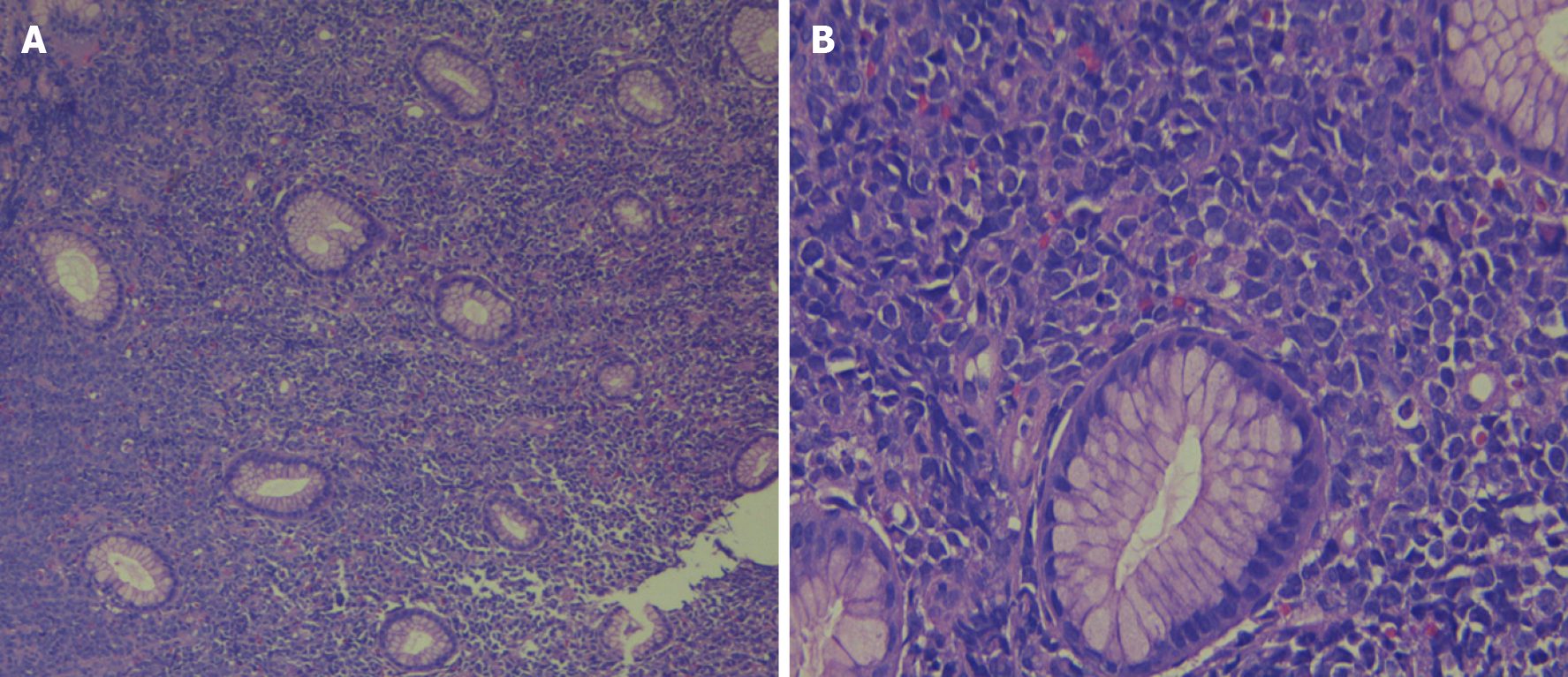
Figure 2 Hematoxylin-eosin staining of gastric myeloid sarcoma.
A: There is mucosa with diffuse infiltration of monomorphic medium cells (× 10 magnification); B: At higher magnification (× 40 magnification) the cells show eosinophilic cytoplasm and nucleus with fine chromatin (blast).
- Citation: Rioja P, Macetas J, Luna-Abanto J, Tirado-Hurtado I, Enriquez DJ. Gastric myeloid sarcoma: A case report. World J Clin Oncol 2021; 12(10): 960-965
- URL: https://www.wjgnet.com/2218-4333/full/v12/i10/960.htm
- DOI: https://dx.doi.org/10.5306/wjco.v12.i10.960


