Copyright
©The Author(s) 2021.
World J Clin Oncol. Oct 24, 2021; 12(10): 868-881
Published online Oct 24, 2021. doi: 10.5306/wjco.v12.i10.868
Published online Oct 24, 2021. doi: 10.5306/wjco.v12.i10.868
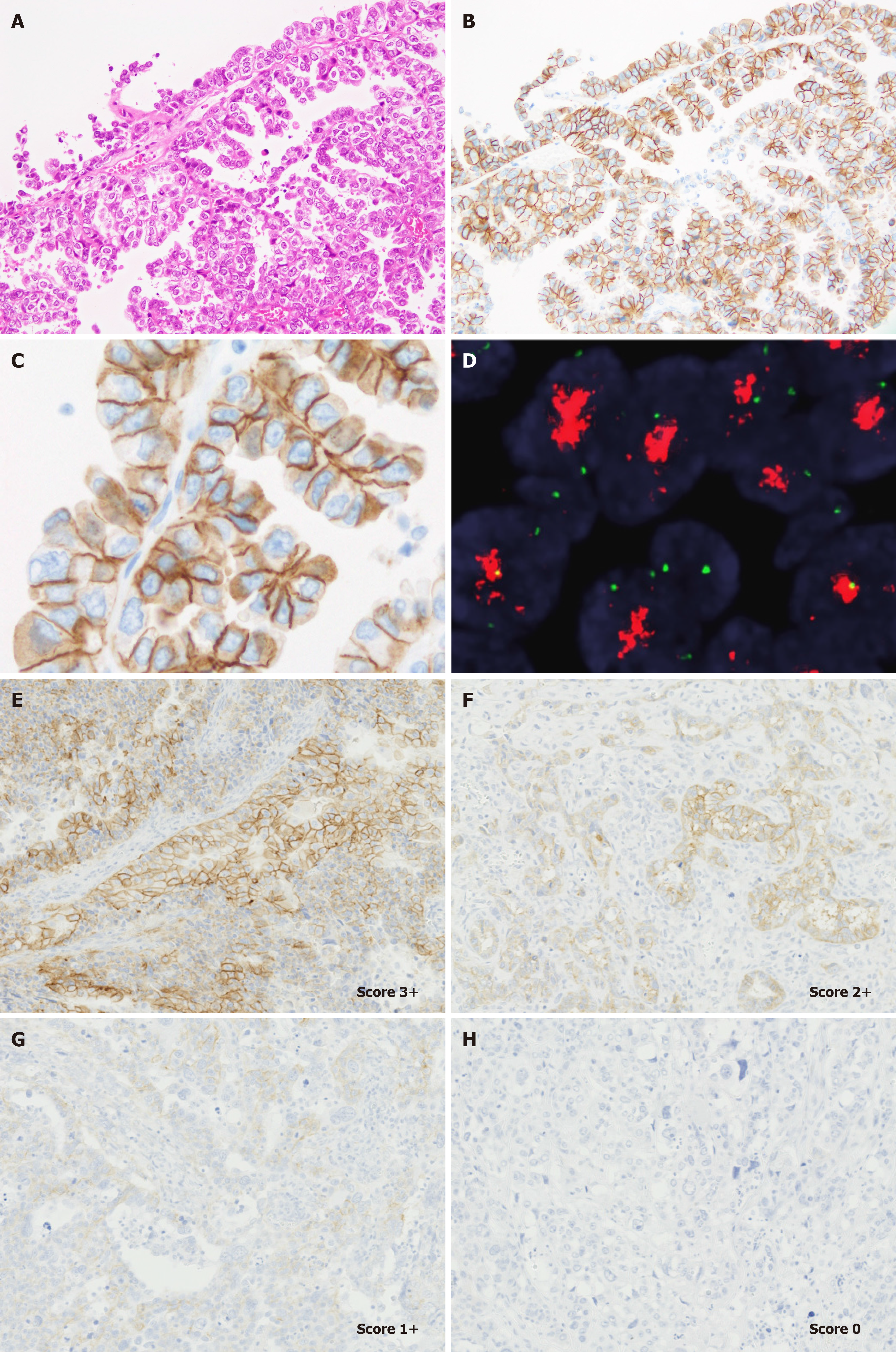
Figure 2 Representative staining patterns of human epidermal growth factor receptor 2 and fluorescent in situ hybridization results in uterine carcinosarcoma.
Serous-like carcinoma component (A) shows diffuse strong membranous staining of Human epidermal growth factor receptor 2 (HER2) (B). Most tumor cells show lateral or basolateral membranous staining patterns (C). HER2 amplification observed in these tumor cells using fluorescent in situ hybridization (D). Representative images of HER2 score according to ASCO/CAP gastric cancer criteria. Score 3+ (E), score 2+ (F), score 1+ (G), and score 0 (H) (× 200). A (H&E, × 100); B, E-G, and H (HER2, × 100); C (HER2, × 400). FISH: Fluorescent in situ hybridization; H&E: Hematoxylin and eosin.
- Citation: Saito A, Yoshida H, Nishikawa T, Yonemori K. Human epidermal growth factor receptor 2 targeted therapy in endometrial cancer: Clinical and pathological perspectives. World J Clin Oncol 2021; 12(10): 868-881
- URL: https://www.wjgnet.com/2218-4333/full/v12/i10/868.htm
- DOI: https://dx.doi.org/10.5306/wjco.v12.i10.868


