Copyright
©The Author(s) 2020.
World J Clin Oncol. Jul 24, 2020; 11(7): 412-427
Published online Jul 24, 2020. doi: 10.5306/wjco.v11.i7.412
Published online Jul 24, 2020. doi: 10.5306/wjco.v11.i7.412
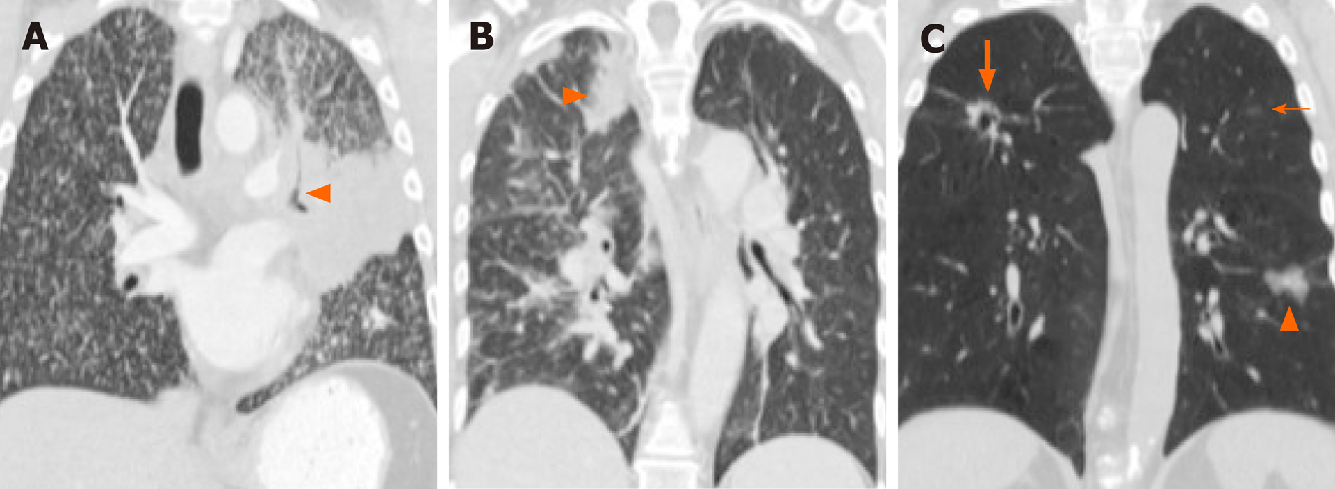
Figure 4 Imaging features of the primary lung tumor and patterns of lung metastases in non-small cell lung cancer with driver mutations.
A: Computed tomography (CT) of a patient with EGFR-mutant non-small cell lung cancer (NSCLC) shows a dominant central left upper lobe mass with diffuse “miliary” type metastases bilaterally. Note the “consolidative” appearance of the dominant mass with air bronchograms (arrowhead), which have also been associated with EGFR-mutant NSCLC; B: CT of a patient with ALK-rearranged NSCLC shows a solid dominant peripheral right upper lobe mass (arrowhead) with nodular thickening of interstitium and ground glass opacities consistent with lymphangitic carcinomatosis, which have been associated with NSCLC with either ALK or ROS1 rearrangements; C: CT of a patient with NSCLC with MET exon 14 skipping mutation shows a part-cystic, part-solid nodule in the right upper lobe (thick arrow) with an additional ground glass nodule in the left lower lobe (arrowhead) and a smaller ground glass nodule in the left upper lobe (thin arrow), consistent with synchronous multifocal lung cancers.
- Citation: Mendoza DP, Piotrowska Z, Lennerz JK, Digumarthy SR. Role of imaging biomarkers in mutation-driven non-small cell lung cancer. World J Clin Oncol 2020; 11(7): 412-427
- URL: https://www.wjgnet.com/2218-4333/full/v11/i7/412.htm
- DOI: https://dx.doi.org/10.5306/wjco.v11.i7.412


