Copyright
©The Author(s) 2020.
World J Clin Oncol. Feb 24, 2020; 11(2): 83-90
Published online Feb 24, 2020. doi: 10.5306/wjco.v11.i2.83
Published online Feb 24, 2020. doi: 10.5306/wjco.v11.i2.83
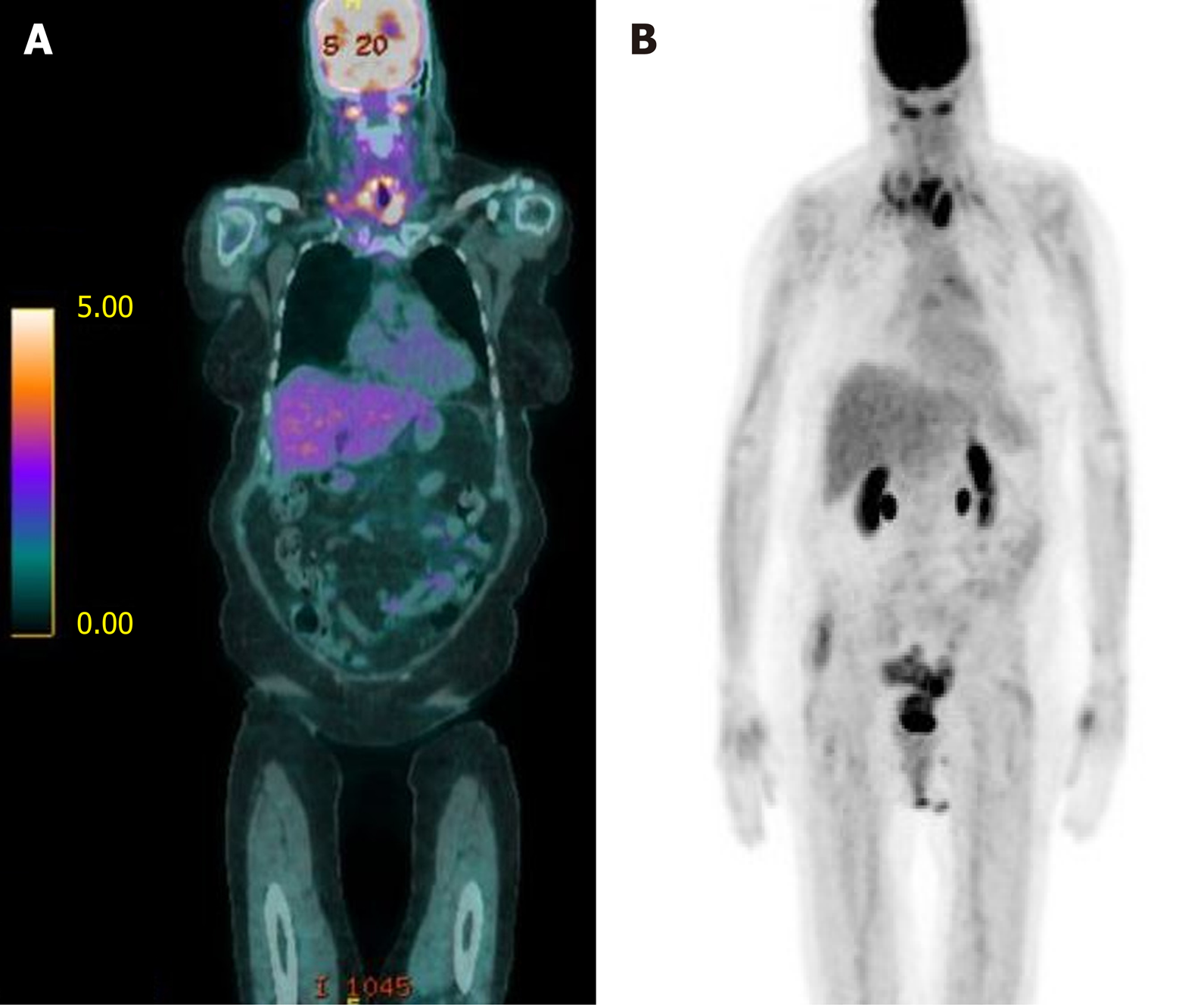
Figure 2 Bilateral lower cervical (SUV 5.
1) and mediastinal (SUV 3.9) adenopathy consistent with metastatic disease. A: Fused coronal positron emission tomography (PET)/ computed tomography (CT) imaging demonstrating an upper esophageal mass (SUV 6.7) as well as bilateral lower cervical adenopathy consistent w/ metastatic disease (SUV 5.1). A few mediastinal lymph nodes are FDG-avid, suspicious for metastatic disease (max SUV 3.9); B: Coronal PET/CT scout with similar uptake noted. PET: Positron Emission Tomography; CT: Computed Tomography.
- Citation: Burns EA, Kasparian S, Khan U, Abdelrahim M. Pancreatic adenocarcinoma with early esophageal metastasis: A case report and review of literature. World J Clin Oncol 2020; 11(2): 83-90
- URL: https://www.wjgnet.com/2218-4333/full/v11/i2/83.htm
- DOI: https://dx.doi.org/10.5306/wjco.v11.i2.83


