Copyright
©The Author(s) 2019.
World J Clin Oncol. Mar 24, 2019; 10(3): 110-135
Published online Mar 24, 2019. doi: 10.5306/wjco.v10.i3.110
Published online Mar 24, 2019. doi: 10.5306/wjco.v10.i3.110
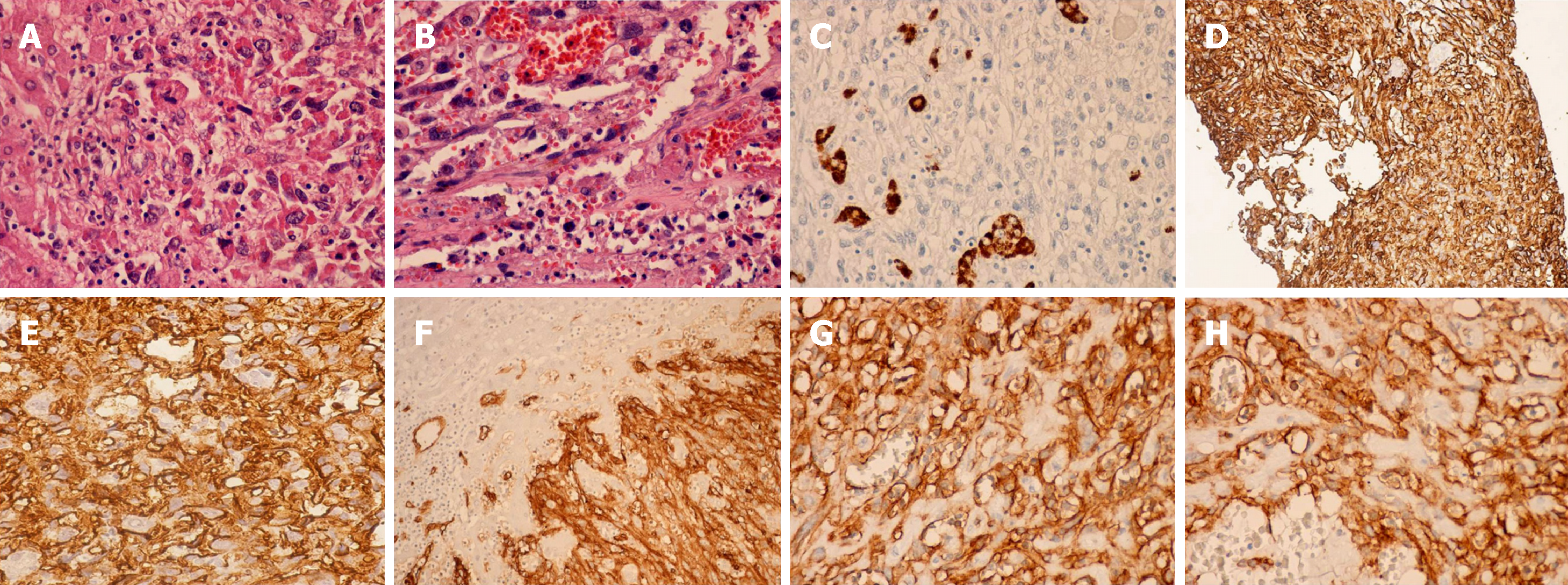
Figure 2 Hepatic angiosarcoma: Pathological findings.
A: Hematoxylin eosin staining, 40× objective; B: Hematoxylin eosin staining, 40× objective, vascular spaces lined by malignant endothelial cells; C: Hep par 1 immunostaining - Hematoxylin eosin background staining, 20× objective; hepatocytes Hep par 1 positive, captive in the tumor mass; D: CD 34 immunostaining, 20× objective, liver biopsy; E: CD 34 immunostaining, 40× objective, liver biopsy (enhanced objective); F: CD34 immunostaining, 20× objective; G: CD31 immunostaining, 20× objective; H: CD31 immunostaining, 40× objective
- Citation: Lazăr DC, Avram MF, Romoșan I, Văcariu V, Goldiș A, Cornianu M. Malignant hepatic vascular tumors in adults: Characteristics, diagnostic difficulties and current management. World J Clin Oncol 2019; 10(3): 110-135
- URL: https://www.wjgnet.com/2218-4333/full/v10/i3/110.htm
- DOI: https://dx.doi.org/10.5306/wjco.v10.i3.110


